PrognoHealth – Corporate Health & Wellness Specialist

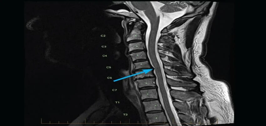
MRI Cervical Spine Magnetic Resonance Imaging (MRI) is a non-invasive diagnostic tool used to create detailed images of internal organs, tissues, and structures of the body. MRI Cervical Spine is a specialized imaging procedure used to evaluate the soft tissues of the neck, including the cervical vertebrae, intervertebral discs, and spinal cord. The procedure is conducted by a trained radiologic technologist, who will help you lie down on a flat table that slides into the MRI machine. The machine uses a strong magnetic field and radio waves to create high-resolution images of the internal structures of the cervical spine. During the procedure, you will need to remain still, and may be asked to hold your breath for short periods of time. Before the test, you will need to remove any metal objects, such as jewelry or watches, as these can interfere with the magnetic field. In some cases, you may be given a contrast agent, such as gadolinium, through an intravenous (IV) injection to enhance the visibility of certain structures. There are various symptoms and conditions that may warrant an MRI Cervical Spine, including neck pain, numbness or tingling in the arms or hands, weakness in the arms or hands, and suspected spinal cord or nerve damage. Additionally, an MRI Cervical Spine may be ordered as part of a routine health checkup or wellness program to screen for any underlying conditions that may not be detected through other diagnostic tests. The length of time for an MRI Cervical Spine can vary, depending on the complexity of the images required, but typically takes around 30-60 minutes. After the test, the images will be analyzed by a radiologist, who will interpret the results and provide a report to your healthcare provider. The results of an MRI Cervical Spine can provide detailed information about the structures and tissues of the cervical spine, including any abnormalities or injuries. The images can be used to diagnose a range of conditions, such as herniated discs, spinal stenosis, or tumors, and can help guide treatment decisions. In addition to its diagnostic uses, an MRI Cervical Spine can also be used in corporate health and wellness programs to screen for underlying conditions that may affect an employee’s health and wellbeing. By identifying potential health issues early, employers can help their employees get the care they need and improve overall productivity and job satisfaction. To prepare for an MRI Cervical Spine, you should inform your healthcare provider if you have any metal implants or devices in your body, as these may be affected by the magnetic field. You may also be asked to fast for a period of time before the test, and should avoid wearing any metal objects, such as jewelry or watches. In summary, MRI Cervical Spine is a non-invasive diagnostic tool that uses magnetic fields and radio waves to create high-resolution images of the internal structures of the neck. It is a safe and effective way to diagnose a range of conditions, including herniated discs, spinal stenosis, and tumors. The test preparation typically involves removing metal objects and, in some cases, receiving a contrast agent through an IV injection. The procedure can take 30-60 minutes, and the results are interpreted by a radiologist who provides a report to your healthcare provider. MRI Cervical Spine can be used in corporate health and wellness programs to screen for underlying conditions that may affect an employee’s health and wellbeing, and help identify potential health issues early. MRI of the Cervical Spine: Magnetic Resonance Imaging (MRI) of the cervical spine is a specialised imaging test that provides detailed pictures of the structures in the neck region, including the vertebrae, spinal cord, intervertebral discs, and surrounding soft tissues. This non-invasive procedure helps diagnose and monitor various conditions affecting the cervical spine. Why is a Cervical Spine MRI Done? A cervical spine MRI is performed for several reasons: 1. Neurological Symptoms:o Neck Pain: Persistent or unexplained pain.o Radiculopathy: Pain radiating to the shoulders or arms, often due to nerve compression.o Myelopathy: Symptoms suggesting spinal cord compression, such as numbness, weakness, or coordination problems in the arms and legs.2. Suspected Structural Abnormalities:o Herniated Discs: Protrusion of intervertebral disc material pressing on nerves or the spinal cord.o Degenerative Disc Disease: Wear and tear of the discs causing pain or stiffness.o Spinal Stenosis: Narrowing of the spinal canal leading to nerve or spinal cord compression.o Spondylosis: Age-related changes in the vertebrae and discs.o Spinal Injuries: Fractures, ligament tears, or other trauma-related changes.3. Tumors and Infections:o Spinal Tumors: Both benign and malignant tumors affecting the spinal column or cord.o Infections: Such as osteomyelitis (bone infection) or discitis (disc infection).4. Autoimmune and Inflammatory Disorders:o Conditions like rheumatoid arthritis or ankylosing spondylitis affecting the cervical spine.5. Post-Surgical Evaluation:o Monitoring recovery or complications after spinal surgery. Anatomy of the Cervical Spine The cervical spine consists of seven vertebrae (C1 to C7) that support the skull and protect the spinal cord. Key structures include: • Vertebrae: The bony segments forming the spine.• Intervertebral Discs: Cushions between the vertebrae that absorb shock.• Spinal Cord: The bundle of nerves running through the spinal canal.• Nerve Roots: Branches of nerves exiting the spinal cord through openings in the vertebrae.• Ligaments and Muscles: Supporting structures that provide stability and movement. Types of MRI Sequences Used for the Cervical Spine Different MRI sequences highlight various tissues and abnormalities: 1. T1-Weighted Imaging:o Good for anatomical details.o Fat appears bright, and fluid appears dark.2. T2-Weighted Imaging:o Excellent for identifying fluid and inflammation.o Fluid and pathology (like a herniated disc) appear bright.3. STIR (Short Tau Inversion Recovery):o Similar to T2 but with suppressed fat signals.o Ideal for detecting edema and inflammation.4. Post-Contrast Imaging:o Uses gadolinium contrast to enhance visualisation of blood vessels, inflammation, and tumors.5. Diffusion-Weighted Imaging (DWI):o Detects changes in the diffusion of water molecules, useful for early detection of spinal cord injury or ischemia. Preparation for a Cervical Spine MRI 1. Safety Screening:o Ensure no contraindications such as metallic implants, pacemakers, or other devices incompatible
Read MoreTop rated products
-
 Full Body Health Checkup I
Original price was: ₹3,000.00.₹1,770.00Current price is: ₹1,770.00.
Full Body Health Checkup I
Original price was: ₹3,000.00.₹1,770.00Current price is: ₹1,770.00.
-
 Young Life Basic
Original price was: ₹3,000.00.₹1,350.00Current price is: ₹1,350.00.
Young Life Basic
Original price was: ₹3,000.00.₹1,350.00Current price is: ₹1,350.00.
-
 COVID 19 Rapid Antigen Test
Original price was: ₹668.00.₹501.00Current price is: ₹501.00.
COVID 19 Rapid Antigen Test
Original price was: ₹668.00.₹501.00Current price is: ₹501.00.
-
 MFG IND PACK III
Original price was: ₹3,900.00.₹2,145.00Current price is: ₹2,145.00.
MFG IND PACK III
Original price was: ₹3,900.00.₹2,145.00Current price is: ₹2,145.00.
-
 Healthy Life Advance Male
Original price was: ₹15,000.00.₹8,250.00Current price is: ₹8,250.00.
Healthy Life Advance Male
Original price was: ₹15,000.00.₹8,250.00Current price is: ₹8,250.00.