PrognoHealth – Corporate Health & Wellness Specialist

Contact Us
+91 9510650660
Email Us
corpsales@prognohealth.com

Contact Us
+91 9510650660
Email Us
corpsales@prognohealth.com
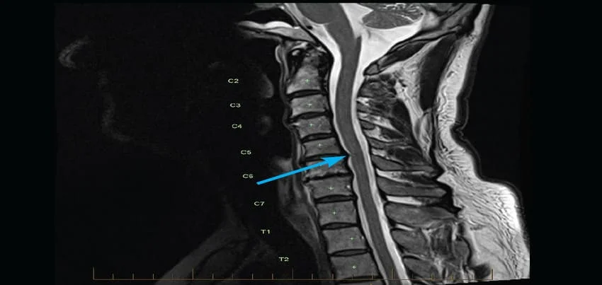
MRI Cervical Spine
Magnetic Resonance Imaging (MRI) is a non-invasive diagnostic tool used to create detailed images of internal organs, tissues, and structures of the body. MRI Cervical Spine is a specialized imaging procedure used to evaluate the soft tissues of the neck, including the cervical vertebrae, intervertebral discs, and spinal cord.
The procedure is conducted by a trained radiologic technologist, who will help you lie down on a flat table that slides into the MRI machine. The machine uses a strong magnetic field and radio waves to create high-resolution images of the internal structures of the cervical spine. During the procedure, you will need to remain still, and may be asked to hold your breath for short periods of time.
Before the test, you will need to remove any metal objects, such as jewelry or watches, as these can interfere with the magnetic field. In some cases, you may be given a contrast agent, such as gadolinium, through an intravenous (IV) injection to enhance the visibility of certain structures.
There are various symptoms and conditions that may warrant an MRI Cervical Spine, including neck pain, numbness or tingling in the arms or hands, weakness in the arms or hands, and suspected spinal cord or nerve damage. Additionally, an MRI Cervical Spine may be ordered as part of a routine health checkup or wellness program to screen for any underlying conditions that may not be detected through other diagnostic tests.
The length of time for an MRI Cervical Spine can vary, depending on the complexity of the images required, but typically takes around 30-60 minutes. After the test, the images will be analyzed by a radiologist, who will interpret the results and provide a report to your healthcare provider.
The results of an MRI Cervical Spine can provide detailed information about the structures and tissues of the cervical spine, including any abnormalities or injuries. The images can be used to diagnose a range of conditions, such as herniated discs, spinal stenosis, or tumors, and can help guide treatment decisions.
In addition to its diagnostic uses, an MRI Cervical Spine can also be used in corporate health and wellness programs to screen for underlying conditions that may affect an employee’s health and wellbeing. By identifying potential health issues early, employers can help their employees get the care they need and improve overall productivity and job satisfaction.
To prepare for an MRI Cervical Spine, you should inform your healthcare provider if you have any metal implants or devices in your body, as these may be affected by the magnetic field. You may also be asked to fast for a period of time before the test, and should avoid wearing any metal objects, such as jewelry or watches.
In summary, MRI Cervical Spine is a non-invasive diagnostic tool that uses magnetic fields and radio waves to create high-resolution images of the internal structures of the neck. It is a safe and effective way to diagnose a range of conditions, including herniated discs, spinal stenosis, and tumors. The test preparation typically involves removing metal objects and, in some cases, receiving a contrast agent through an IV injection. The procedure can take 30-60 minutes, and the results are interpreted by a radiologist who provides a report to your healthcare provider. MRI Cervical Spine can be used in corporate health and wellness programs to screen for underlying conditions that may affect an employee’s health and wellbeing, and help identify potential health issues early.
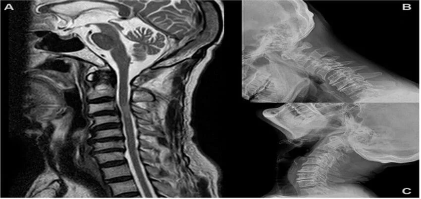
MRI of the Cervical Spine:
Magnetic Resonance Imaging (MRI) of the cervical spine is a specialised imaging test that provides detailed pictures of the structures in the neck region, including the vertebrae, spinal cord, intervertebral discs, and surrounding soft tissues. This non-invasive procedure helps diagnose and monitor various conditions affecting the cervical spine.
Why is a Cervical Spine MRI Done?
A cervical spine MRI is performed for several reasons:
1. Neurological Symptoms:
o Neck Pain: Persistent or unexplained pain.
o Radiculopathy: Pain radiating to the shoulders or arms, often due to nerve compression.
o Myelopathy: Symptoms suggesting spinal cord compression, such as numbness, weakness, or coordination problems in the arms and legs.
2. Suspected Structural Abnormalities:
o Herniated Discs: Protrusion of intervertebral disc material pressing on nerves or the spinal cord.
o Degenerative Disc Disease: Wear and tear of the discs causing pain or stiffness.
o Spinal Stenosis: Narrowing of the spinal canal leading to nerve or spinal cord compression.
o Spondylosis: Age-related changes in the vertebrae and discs.
o Spinal Injuries: Fractures, ligament tears, or other trauma-related changes.
3. Tumors and Infections:
o Spinal Tumors: Both benign and malignant tumors affecting the spinal column or cord.
o Infections: Such as osteomyelitis (bone infection) or discitis (disc infection).
4. Autoimmune and Inflammatory Disorders:
o Conditions like rheumatoid arthritis or ankylosing spondylitis affecting the cervical spine.
5. Post-Surgical Evaluation:
o Monitoring recovery or complications after spinal surgery.
Anatomy of the Cervical Spine
The cervical spine consists of seven vertebrae (C1 to C7) that support the skull and protect the spinal cord. Key structures include:
• Vertebrae: The bony segments forming the spine.
• Intervertebral Discs: Cushions between the vertebrae that absorb shock.
• Spinal Cord: The bundle of nerves running through the spinal canal.
• Nerve Roots: Branches of nerves exiting the spinal cord through openings in the vertebrae.
• Ligaments and Muscles: Supporting structures that provide stability and movement.
Types of MRI Sequences Used for the Cervical Spine
Different MRI sequences highlight various tissues and abnormalities:
1. T1-Weighted Imaging:
o Good for anatomical details.
o Fat appears bright, and fluid appears dark.
2. T2-Weighted Imaging:
o Excellent for identifying fluid and inflammation.
o Fluid and pathology (like a herniated disc) appear bright.
3. STIR (Short Tau Inversion Recovery):
o Similar to T2 but with suppressed fat signals.
o Ideal for detecting edema and inflammation.
4. Post-Contrast Imaging:
o Uses gadolinium contrast to enhance visualisation of blood vessels, inflammation, and tumors.
5. Diffusion-Weighted Imaging (DWI):
o Detects changes in the diffusion of water molecules, useful for early detection of spinal cord injury or ischemia.
Preparation for a Cervical Spine MRI
1. Safety Screening:
o Ensure no contraindications such as metallic implants, pacemakers, or other devices incompatible with MRI.
o Inform your healthcare provider about any prior surgeries, tattoos, or metal fragments in the body.
2. Clothing and Accessories:
o Wear comfortable, metal-free clothing.
o Remove all jewellery, watches, and accessories.
3. Eating and Drinking:
o No specific restrictions, but follow any instructions given by your healthcare provider.
4. Claustrophobia:
o Inform the technologist if you have claustrophobia. Options like open MRI machines or sedation can be considered.
5. Contrast Considerations:
o If contrast material is to be used, notify your provider about any kidney issues or allergies to gadolinium-based agents.
The Procedure
1. Positioning:
o You will lie on your back on a motorized table that slides into the MRI scanner.
o Your head and neck will be positioned and secured to minimise movement and ensure high-quality images.
2. Imaging:
o The scan typically takes 30-60 minutes.
o You will need to remain still to avoid blurring the images.
o You will hear a series of loud thumping or buzzing noises; earplugs or headphones are often provided.
3. Communication:
o You can speak with the MRI technologist through an intercom if you need assistance or have concerns during the scan.
4. Contrast Injection:
o If contrast is required, it will be injected through an IV partway through the scan.
Interpretation of MRI Results
The MRI results are analyzed by a radiologist, who will provide a detailed report highlighting any abnormalities, including:
1. Disc Issues:
o Herniated Discs: Bulging or ruptured discs pressing on nerves or the spinal cord.
o Degenerative Disc Disease: Thinning or degeneration of the discs.
2. Bone Abnormalities:
o Spondylosis: Arthritis-related changes in the vertebrae.
o Fractures: Breaks or cracks in the vertebrae.
3. Spinal Cord and Nerve Issues:
o Spinal Stenosis: Narrowing of the spinal canal causing compression.
o Myelopathy: Damage or compression of the spinal cord.
o Radiculopathy: Compression of nerve roots leading to pain or numbness in the arms.
4. Soft Tissue Changes:
o Ligament and Muscle Injuries: Tears or strains.
o Tumors: Benign or malignant growths affecting the spinal structures.
5. Inflammatory or Infectious Conditions:
o Discitis or Osteomyelitis: Infections affecting the discs or vertebrae.
o Autoimmune Disorders: Inflammatory changes associated with conditions like rheumatoid arthritis.
Advantages of Cervical Spine MRI
1. High-Resolution Images:
o Detailed visualization of soft tissues, spinal cord, and intervertebral discs.
2. Non-Invasive and Safe:
o No exposure to ionizing radiation.
3. Comprehensive Evaluation:
o Can assess a wide range of conditions affecting the cervical spine.
4. Contrast Enhancement:
o Provides better visualization of blood flow and pathology when contrast is used.
Limitations and Considerations
1. Cost and Availability:
o MRI is more expensive and less readily available compared to other imaging modalities like X-ray or CT scans.
2. Duration and Patient Comfort:
o Longer procedure times and potential discomfort from staying still in a confined space.
3. Contraindications:
o Not suitable for patients with certain metallic implants or severe claustrophobia.
4. Gadolinium Risks:
o Rare but potential risks with gadolinium contrast, especially in patients with kidney issues.
Summary :
MRI of the cervical spine is an essential diagnostic tool for evaluating a wide range of neck and spinal conditions. It provides detailed, high-resolution images and is invaluable for diagnosing, monitoring, and planning treatment for various cervical spine disorders. Proper preparation and understanding of the procedure help ensure accurate results and optimal care.
References :
• RadiologyInfo.org: MRI of the Spine
• American Society of Neuroradiology: Cervical Spine MRI
Login
0 Comments
Inline Feedbacks
View all comments
Blog Categories
Top rated products
-
 FB Health Checkup I
Rated 5.00 out of 5
FB Health Checkup I
Rated 5.00 out of 5₹3,000.00Original price was: ₹3,000.00.₹1,770.00Current price is: ₹1,770.00. -
Progno Health III
Rated 0 out of 5
₹3,900.00Original price was: ₹3,900.00.₹2,145.00Current price is: ₹2,145.00. -
Healthy Diva
Rated 0 out of 5
₹9,000.00Original price was: ₹9,000.00.₹5,850.00Current price is: ₹5,850.00. -
 Advance Health Check Plus -M/F -98 Parameters
Rated 0 out of 5₹1,999.00
Advance Health Check Plus -M/F -98 Parameters
Rated 0 out of 5₹1,999.00 -
IT & ITES PACK II
Rated 0 out of 5
₹12,000.00Original price was: ₹12,000.00.₹6,600.00Current price is: ₹6,600.00.
About Us
Progno Health is a Corporate Health & Wellness Specialist providing services to Pan India. We offer Pre-employment Health Checkup Packages, Annual Health Checkup Packages, Executive Health Checkup Packages, Occupational Health Checkup Packages, and other Health & Wellness Services.
