PrognoHealth – Corporate Health & Wellness Specialist

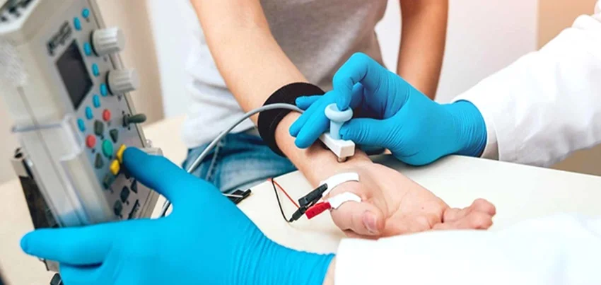
Electromyography (EMG) Electromyography (EMG) is a diagnostic test used to evaluate the electrical activity of muscles and nerves. The test is conducted by inserting a small needle electrode into the muscle tissue, which measures the electrical signals produced by the muscle. These signals are then recorded and analyzed by a machine to provide information about the functioning of the muscle and nerve tissue. Test Preparation:There is typically no special preparation required for an EMG. However, patients may be asked to avoid caffeine or certain medications prior to the test. They may also be asked to wear loose clothing that allows easy access to the muscles being tested. Common Symptoms for ordering the test:An EMG may be ordered for a variety of reasons, such as: Muscle weakness or atrophyNumbness or tingling in the extremitiesPain or cramping in the musclesSuspected nerve damage or injurySuspected muscle or nerve disorders, such as muscular dystrophy or multiple sclerosis Time taken for the test:The test itself typically takes between 30 minutes to an hour to complete, depending on the number of muscles being tested. In some cases, the test may be prolonged for several hours or even days if a patient is being monitored for a specific condition. Results Interpretations:The results of an EMG are interpreted by a trained healthcare professional, such as a neurologist or physiatrist. The results may show abnormalities in muscle or nerve activity, such as decreased or increased muscle activity, or abnormal patterns of electrical activity. In some cases, the results may be inconclusive or require further testing. The healthcare professional will use the results of the EMG, along with other medical information and test results, to make a diagnosis and develop a treatment plan for the patient. Health checkups, wellness, and corporate health are all areas where EMG testing may be used. Regular health checkups can help detect early signs of muscle or nerve disorders, which may benefit from early intervention and treatment. Wellness programs may incorporate EMG testing as a way to assess muscle function and identify areas for improvement. In the corporate health setting, EMG testing may be used to evaluate workers who perform repetitive or strenuous tasks, such as assembly line workers or construction workers. By identifying early signs of muscle or nerve damage, employers can take steps to prevent further injury and promote worker safety. In conclusion, electromyography is a diagnostic test used to evaluate the electrical activity of muscles and nerves. The test is conducted by inserting a small needle electrode into the muscle tissue, which measures the electrical signals produced by the muscle. EMG testing may be used to diagnose muscle and nerve disorders, evaluate muscle function, and monitor the effects of treatment. The test typically takes between 30 minutes to an hour to complete, depending on the number of muscles being tested. Results are interpreted by a trained healthcare professional and used to make a diagnosis and develop a treatment plan. EMG testing may be incorporated into health checkups, wellness programs, and corporate health initiatives as a way to promote early detection and prevention of muscle and nerve disorders.
Read More
Guillain-Barré Syndrome Guillain-Barré syndrome (GBS) is a rare disorder that affects the nervous system and can cause muscle weakness, tingling sensations, and even paralysis. It is caused by an abnormal immune response that attacks the peripheral nerves, which are the nerves that control muscle movement and sensation in the arms and legs. Symptoms of GBS can vary depending on the person, but common symptoms include muscle weakness, tingling sensations, and loss of reflexes in the arms and legs. In some cases, GBS can also cause difficulty breathing, which can be life-threatening. Symptoms usually start in the feet and legs, and then progress upward. The onset of symptoms may be sudden or gradual. Diagnosis of GBS is based on the patient’s symptoms, medical history, and lab tests. A nerve conduction study and an electromyography (EMG) can be used to measure the electrical activity in the muscles and nerves, which can help to confirm the diagnosis. A lumbar puncture (spinal tap) may also be done to check for antibodies in the cerebrospinal fluid, which can indicate GBS. Treatment of GBS typically involves the use of immunoglobulin (IVIg) or plasma exchange (PE). IVIg is a treatment that uses antibodies from healthy donors to help reduce inflammation in the nerves, while PE is a procedure that removes the plasma from the blood and replaces it with fresh plasma. In addition, supportive care such as physical therapy, occupational therapy, and breathing support may be needed to help patients recover from muscle weakness and paralysis. The cause of GBS is not well understood, but it is thought to be triggered by an infection or other underlying medical condition. Certain viral and bacterial infections have been linked to the development of GBS, such as Campylobacter jejuni, cytomegalovirus, Epstein-Barr virus, and HIV. Preventing GBS from occurring is difficult, as the exact cause is not yet known. However, there are certain things that can be done to reduce the risk of developing the condition, such as getting vaccinated against certain infections, and seeking treatment for any underlying medical conditions. Regular check-ups and annual health screenings are important for early detection and treatment of GBS. People who are at a higher risk of developing GBS, such as those with an underlying medical condition or who have recently had an infection, should consider getting checked more frequently. In terms of diet and exercise, there is no specific diet or exercise regimen that can prevent the development of GBS. However, maintaining a healthy lifestyle with a balanced diet and regular physical activity can help to boost the immune system and reduce the risk of developing certain infections that may trigger GBS. Corporate health and wellness programs can play a key role in preventing GBS by encouraging regular check-ups and annual health screenings, providing education on the importance of getting vaccinated and seeking treatment for underlying medical conditions. It can also provide information on how to maintain a healthy lifestyle with a balanced diet and regular physical activity, which can help to boost the immune system. In conclusion, Guillain-Barré syndrome (GBS) is a rare disorder that affects the nervous system and can cause muscle weakness, tingling sensations, and even paralysis. The cause is not well understood, but it is thought to be triggered by an infection or other underlying medical condition. Diagnosis is based on the patient’s symptoms, medical history, and lab tests. Treatment typically involves the use of immunoglobulin (IVIg) or plasma exchange (PE), and supportive care. Preventing GBS from occurring is difficult.
Read More
MRI of the Lumbar and Sacral Spine Magnetic Resonance Imaging (MRI) of the Lumbar and Sacral Spine is a non-invasive diagnostic test that uses a powerful magnetic field, radio waves, and a computer to produce detailed images of the lower back and pelvis. The test is conducted to evaluate a range of conditions affecting the lumbar and sacral spine, including injuries, disc herniation, spinal stenosis, tumors, and other abnormalities. Test Conducted: During an MRI of the Lumbar and Sacral Spine, the patient lies on a table that slides into a cylindrical machine that houses the MRI scanner. The scanner uses a magnetic field and radio waves to create images of the lumbar and sacral spine. The test typically takes between 30 and 60 minutes to complete, depending on the complexity of the exam and the patient’s ability to remain still during the procedure. Test Preparation: For an MRI of the Lumbar and Sacral Spine, patients should wear comfortable, loose-fitting clothing without metal zippers or buttons. The patient will be asked to remove any metal objects, such as jewelry, watches, or belts. In some cases, the patient may need to fast for a few hours before the exam, depending on the reason for the test. Common Symptoms for Ordering the Test: MRI of the Lumbar and Sacral Spine may be ordered by a physician if a patient has symptoms such as lower back pain, leg pain, numbness or tingling in the legs or feet, weakness in the legs or feet, or difficulty standing or walking. The test can help diagnose a range of conditions affecting the lumbar and sacral spine, including injuries, disc herniation, spinal stenosis, tumors, and other abnormalities. Time Taken for the Test and Interpretation of Results: An MRI of the Lumbar and Sacral Spine typically takes between 30 and 60 minutes to complete, and patients can return to their normal activities immediately after the test. The images produced by the MRI are examined by a radiologist, who will interpret the results and provide a report to the ordering physician. The physician will then review the results with the patient and determine the appropriate course of treatment based on the findings.MRI of the Lumbar and Sacral Spine is an important tool for diagnosing and treating a range of conditions affecting the lower back and pelvis, and can be used in regular health checkups and corporate wellness programs. Early detection of lumbar and sacral spine problems is critical to maintaining good health and wellness. In addition, some corporate health programs offer wellness screenings that include lower back exams, and an MRI of the Lumbar and Sacral Spine may be ordered as part of a comprehensive wellness evaluation. In conclusion, MRI of the Lumbar and Sacral Spine is a valuable diagnostic tool that can help detect a range of lower back and pelvis conditions that can affect overall health and wellness. The test is non-invasive and typically takes between 30 and 60 minutes to complete, with some preparation required such as removing metal objects and fasting for a few hours before the exam. The results are interpreted by a radiologist and reviewed by the ordering physician, who will determine the appropriate course of treatment based on the findings. Regular health checkups and corporate wellness programs can also incorporate lower back exams, including MRI of the Lumbar and Sacral Spine, to help detect and manage lumbar and sacral spine problems.
Read More
What Is A Full Body Health Checkup & Why It Is Important? We All Know The Famous Adage: Prevention Is Better Than Cure People need to take care of their health right from a young age, so they don’t undergo serious health problems later in life.
Read More
CT Scan Spiral Angio Brain CT scan Spiral Angio Brain is a non-invasive diagnostic imaging test that helps in assessing the blood vessels of the brain. It is primarily used to detect any abnormalities in the brain’s blood vessels, such as blockages, aneurysms, or other malformations. This test uses a combination of X-rays and computer technology to produce detailed images of the brain’s blood vessels. The CT Scan Spiral Angio Brain is conducted with the help of a CT machine, which is a large, doughnut-shaped device. The patient will lie down on a table that slides into the CT machine. The table will move slowly through the machine, and the X-ray beams will rotate around the patient’s body. The CT machine will capture detailed images of the brain’s blood vessels from various angles. The process usually takes around 30 to 45 minutes to complete. There is no specific preparation required for the CT Scan Spiral Angio Brain. However, the patient may be asked to avoid eating or drinking anything for several hours before the test. In some cases, a contrast dye may be injected into the patient’s veins before the test to help the blood vessels appear more clearly in the images. If a contrast dye is used, the patient will need to inform their doctor if they have any allergies or kidney problems. Some common symptoms that may lead to the ordering of a CT Scan Spiral Angio Brain include headaches, dizziness, vision problems, weakness or numbness in the arms or legs, seizures, or a family history of aneurysms or other vascular abnormalities. In addition, this test may be used to evaluate patients who have had a stroke or to assess the blood flow in the brain before surgery. After the test, the images produced by the CT Scan Spiral Angio Brain are interpreted by a radiologist, who is a doctor specialized in diagnostic imaging. The radiologist will review the images and provide a report to the referring physician. The results of the test can help the doctor make a diagnosis and determine the best course of treatment. Overall, the CT Scan Spiral Angio Brain is a safe and effective imaging test that can provide important information about the blood vessels in the brain. It is a valuable tool for diagnosing and managing a variety of neurological conditions. As with any medical procedure, patients should discuss the risks and benefits of this test with their doctor before undergoing the test.
Read More
CT Scan of the Thorax A CT (computed tomography) scan of the thorax, also known as a chest CT scan, is a non-invasive medical imaging test that produces detailed images of the chest and surrounding structures. The test is commonly used to diagnose and evaluate a wide range of lung and chest conditions. In this article, we will discuss the procedure of the CT Scan Thorax, test preparation, common symptoms for ordering the test, time taken for the test, result interpretation, and its importance in corporate health wellness packages. Test Procedure : During a CT Scan Thorax, the patient lies on a table that slides through a large, doughnut-shaped machine that rotates around the patient’s body. The machine takes multiple images of the chest, including the lungs, heart, blood vessels, and other surrounding structures. The images are then processed by a computer to create detailed, three-dimensional images of the chest. The entire procedure typically takes between 10 to 30 minutes. Test Preparation : Before a CT Scan Thorax, patients may be asked to refrain from eating or drinking for a few hours, especially if they will receive contrast dye. Contrast dye is a substance that enhances the visibility of certain structures in the body during imaging. Patients should also inform their doctor of any medications they are taking, any allergies they have, and any previous surgeries or medical procedures. In some cases, patients may be asked to wear a hospital gown or remove any metal objects, such as jewelry or piercings. Common Symptoms for Ordering the Test : A CT Scan Thorax may be ordered by a doctor to investigate the following symptoms or conditions:Persistent cough or chest painShortness of breath or difficulty breathingUnexplained weight lossFatigue or weaknessSuspected lung or chest infectionSuspected blood clot in the lungEvaluation of lung nodules or masses Time Taken for the Test and its Results Interpretation : The CT Scan Thorax usually takes between 10 to 30 minutes to complete. After the test, a radiologist will interpret the images and provide a report to the referring physician. The report will include information about the condition of the lungs, heart, and other structures in the chest, including any abnormalities such as masses, nodules, or signs of inflammation. Importance in Corporate Health Wellness Packages : CT Scan Thorax is a critical diagnostic tool that can detect and diagnose a wide range of lung and chest conditions. Early detection and treatment of these conditions can help prevent complications, reduce the need for more invasive procedures, and ultimately lead to better health outcomes. By offering CT Scan Thorax as part of corporate health wellness packages, companies can help their employees detect and diagnose these conditions early, leading to better overall health and productivity. In conclusion, CT Scan Thorax is a non-invasive medical imaging test used to diagnose and evaluate a wide range of lung and chest conditions. Patients do not need to prepare significantly for this test, but they should inform their doctor of any medications they are taking, any allergies they have, and any previous surgeries or medical procedures. Results of the test are interpreted by a radiologist and provided to the referring physician. Companies can offer CT Scan Thorax as part of their corporate wellness packages to help their employees detect and diagnose these conditions early and promote better overall health and productivity. CT scan of the thorax A CT scan of the thorax, also known as a chest CT scan, is a diagnostic imaging procedure that provides detailed pictures of the structures within the chest, including the lungs, heart, blood vessels, and bones. This scan is invaluable for diagnosing a variety of conditions related to the respiratory and cardiovascular systems as well as other structures in the chest. Overview of a Thoracic CT Scan • CT Scan : Computed Tomography (CT) uses X-rays to create cross-sectional images of the body, offering more detailed information than standard X-rays.• Thorax : The thorax is the part of the body between the neck and the abdomen. It includes vital structures such as the lungs, heart, esophagus, trachea, and major blood vessels. Indications for a Thoracic CT Scan A thoracic CT scan is performed for various reasons, including: 1. Evaluation of Symptoms 😮 Persistent cough.o Shortness of breath.o Chest pain.o Hemoptysis (coughing up blood).2. Detection and Assessment of Conditions 😮 Lung Diseases : Such as pneumonia, tuberculosis, interstitial lung disease, and chronic obstructive pulmonary disease (COPD).o Tumors : Including lung cancer, metastatic disease, and lymphomas.o Infections : To identify the presence and extent of infections like abscesses.o Pulmonary Embolism : Blood clots in the lungs.o Aortic Aneurysms : Enlargement of the aorta.o Pleural Conditions : Such as pleural effusion (fluid around the lungs) or pneumothorax (collapsed lung).o Congenital Abnormalities : Birth defects affecting the heart, lungs, or chest structures.3. Preoperative Assessment 😮 Planning for surgeries involving the chest organs.o Guiding biopsies or other interventional procedures.4. Monitoring of Chronic Conditions 😮 Following up on known tumors or chronic lung diseases.o Evaluating the effectiveness of ongoing treatments. Preparation for the Scan 1. Dietary Restrictions : Usually, no fasting is required unless a contrast dye is used.2. Contrast Use 😮 Without Contrast : No special preparation is needed.o With Contrast : You may be asked to avoid eating for a few hours before the scan. Inform your doctor if you have any allergies or kidney issues, as these can affect the use of contrast dye.3. Clothing and Jewelry : Remove any metal objects, such as jewelry, that might interfere with the imaging.4. Medical History : Inform the technician if you have any medical conditions, are pregnant, or have had prior reactions to contrast materials. Procedure 1. Positioning : You will lie flat on a motorized table. You may be asked to raise your arms above your head.2. Scanning : The table will slide into the CT scanner, a large doughnut-shaped machine. The scanner rotates around your body, capturing multiple X-ray images.3. Breath Holding : You may be instructed to hold your breath
Read More
CT Scan Spiral Brain CT scan spiral brain is a medical test that is used to produce highly detailed images of the brain using X-rays. It is also known as a helical CT scan or a spiral CT scan. The procedure is fast, efficient and relatively safe, and it is commonly used to detect various types of brain conditions, such as tumors, bleeding or swelling. The CT scan spiral brain is conducted using a large, circular machine that rotates around the patient’s head while they lie on a table. Unlike traditional CT scans, spiral CT scans are continuous and take images of the brain from all angles, creating a 3D image of the brain. The machine moves very slowly, which allows for more detailed images to be taken. The procedure is non-invasive and generally painless, and there is usually no preparation required for the patient prior to the test. However, patients may be asked to avoid food and drink for a few hours before the procedure. Patients should also let their doctor know if they are pregnant or have any allergies to medications or contrast agents. The most common symptoms that may require a CT scan spiral brain include headaches, seizures, dizziness, changes in vision or speech, or any other neurological symptoms. Patients who have had an injury to the head or have a family history of neurological conditions may also require a CT scan spiral brain. The procedure takes only a few minutes, and the patient is required to remain still throughout the scan. During the procedure, the patient may be asked to hold their breath for a few seconds while the machine takes the images. After the procedure, the patient can resume their normal activities immediately. The results of the CT scan spiral brain will be interpreted by a radiologist, who will provide a report to the patient’s doctor. The report will indicate any abnormalities in the brain, such as tumors, bleeding, or swelling. The doctor will then discuss the results with the patient and determine the appropriate course of treatment. CT scans of the brain, including spiral CT scans, can be an important part of health checkups, wellness programs, and corporate health programs. They are used to screen for potential brain conditions and ensure early detection and treatment. However, it is important to note that CT scans involve exposure to ionizing radiation, and should only be used when medically necessary. In conclusion, a CT scan spiral brain is a highly effective diagnostic tool for detecting brain conditions, such as tumors, bleeding, or swelling. The procedure is fast, efficient and relatively safe, and is commonly used as part of health checkups, wellness programs, and corporate health programs. If you are experiencing any neurological symptoms or have had an injury to the head, speak with your doctor to determine if a CT scan spiral brain is necessary.
Read More
Dengue, a mosquito-borne viral disease, poses a significant health risk globally, including India. By understanding the origins of this silent threat, we can better equip ourselves with knowledge
Read More
Iron deficiency anemia Iron deficiency anemia is a condition in which the body lacks enough iron to produce enough healthy red blood cells. These cells are responsible for carrying oxygen to the body’s tissues, so a lack of them can lead to fatigue, weakness, and a host of other symptoms. Symptoms of iron deficiency anemia include fatigue, weakness, pale skin, shortness of breath, cold hands and feet, fast heartbeat, and chest pain. In severe cases, the condition can also cause headaches, dizziness, and poor appetite. To diagnose iron deficiency anemia, a doctor will typically perform a physical examination and take a blood sample to measure the levels of hemoglobin, the protein in red blood cells that carries oxygen. The doctor may also order other tests such as a complete blood count (CBC), a ferritin test, and a transferrin test to confirm the diagnosis. Common treatment methods for iron deficiency anemia include taking iron supplements and increasing iron-rich foods in the diet. In some cases, a doctor may also recommend a blood transfusion to increase the number of healthy red blood cells in the body. To prevent iron deficiency anemia from occurring, it is important to maintain a healthy diet that includes plenty of iron-rich foods such as lean red meat, poultry, fish, beans, and leafy greens. Regular exercise can also help to improve the body’s ability to absorb iron. Annual health check-ups and corporate health & wellness programs can also play a role in preventing iron deficiency anemia. These programs can help to identify individuals who may be at risk of developing the condition and provide them with the necessary education and resources to make lifestyle changes to prevent it. Diet and exercise are key preventions of Iron deficiency anemia. Eating a diet that is rich in iron-rich foods, such as lean red meat, poultry, fish, beans, and leafy greens, can help to prevent the condition. Regular exercise can also help to improve the body’s ability to absorb iron. It is also important to maintain a healthy weight, as being overweight or obese can increase the risk of developing iron deficiency anemia. Incorporating annual health check-ups and corporate health & wellness programs can also be very helpful in preventing iron deficiency anemia. These programs can help identify individuals who may be at risk of developing the condition and provide them with the necessary education and resources to make lifestyle changes to prevent it. Overall, iron deficiency anemia is a serious condition that can cause a wide range of symptoms, but it is also preventable. By maintaining a healthy diet, exercising regularly, and participating in annual health check-ups and corporate health & wellness programs, individuals can reduce their risk of developing the condition and enjoy better overall health
Read More
A Glycosylated Hemoglobin (HbA1c) test is a blood test that measures the percentage of hemoglobin that has been coated with glucose (sugar) molecules.
Read MoreTop rated products
-
 Full Body Health Checkup I
Original price was: ₹3,000.00.₹1,770.00Current price is: ₹1,770.00.
Full Body Health Checkup I
Original price was: ₹3,000.00.₹1,770.00Current price is: ₹1,770.00.
-
 MFG IND PACK III
Original price was: ₹3,900.00.₹2,145.00Current price is: ₹2,145.00.
MFG IND PACK III
Original price was: ₹3,900.00.₹2,145.00Current price is: ₹2,145.00.
-
 Healthy Life Advance Male
Original price was: ₹15,000.00.₹8,250.00Current price is: ₹8,250.00.
Healthy Life Advance Male
Original price was: ₹15,000.00.₹8,250.00Current price is: ₹8,250.00.
-
 HealthGuard Pro -Male – 80 Parameters
₹849.00
HealthGuard Pro -Male – 80 Parameters
₹849.00
-
 Progno Health IV
Original price was: ₹8,000.00.₹4,400.00Current price is: ₹4,400.00.
Progno Health IV
Original price was: ₹8,000.00.₹4,400.00Current price is: ₹4,400.00.