PrognoHealth – Corporate Health & Wellness Specialist


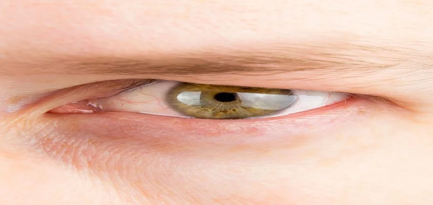
Dermatochalasis Dermatochalasis, also known as excess skin of the upper eyelid, is a common condition that affects people of all ages. It occurs when the skin of the upper eyelid becomes loose and droopy, giving a tired or aged appearance. Symptoms of dermatochalasis include a heavy, drooping appearance of the upper eyelid, difficulty keeping the eyelid open, and even vision impairment. These symptoms can make people feel self-conscious and can also affect their daily activities. The diagnosis of dermatochalasis is usually made through a physical examination by a qualified medical professional. During the examination, the doctor will assess the appearance of the eyelid, as well as any vision impairment or other symptoms that may be present. Common treatment methods for dermatochalasis include blepharoplasty, a surgical procedure that removes excess skin and fat from the upper eyelid. The procedure is usually done on an outpatient basis and requires minimal recovery time. Another treatment option is non-surgical, such as Botox injections to temporarily paralyze the muscle that causes the eyelid to droop. Preventing dermatochalasis from occurring can be done through several methods. One of the most effective ways to prevent it from occurring is through regular annual health check-ups and corporate health & wellness programs. These check-ups can help to identify and address any underlying conditions that may be contributing to the development of dermatochalasis. Additionally, maintaining a healthy diet and regular exercise can also help to prevent dermatochalasis from occurring. A diet that is rich in fruits, vegetables, and lean protein can help to support the overall health of the skin and the body. Regular exercise can also help to keep the skin and muscles toned, which can help to prevent the eyelids from drooping. In summary, Dermatochalasis is a common condition that can affect people of all ages. It is characterized by a heavy, drooping appearance of the upper eyelid and can cause vision impairment and self-consciousness. The condition can be diagnosed through a physical examination and treated through surgery or non-surgical options. To prevent it from occurring, regular annual health check-ups and corporate health & wellness programs, healthy diet, and regular exercise are recommended.
Read More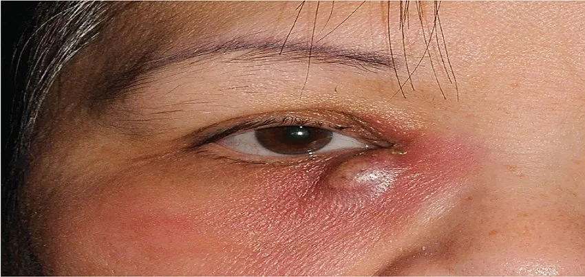
Dacryocystitis Dacryocystitis is an infection of the lacrimal sac, which is located at the inner corner of the eye and is responsible for draining tears from the eye. The condition is characterized by inflammation and blockage of the lacrimal duct, which can lead to the accumulation of pus and the formation of an abscess. Symptoms of dacryocystitis can include pain and swelling in the affected area, redness of the eye, and a thick, green or yellow discharge from the eye. Other symptoms may include fever, fatigue, and a general feeling of malaise. In some cases, the infection may spread to other parts of the face, causing additional symptoms such as a sinus infection or cellulitis. The diagnosis of dacryocystitis is typically made through a physical examination and imaging tests such as a CT scan or MRI. In some cases, a culture of the discharge may also be taken to identify the specific type of bacteria causing the infection. Treatment for dacryocystitis typically involves the use of antibiotics to clear the infection, as well as anti-inflammatory medications to reduce inflammation and pain. In some cases, surgical intervention may be necessary to drain the abscess or remove the affected lacrimal duct. It’s important to note that dacryocystitis can be caused by a variety of factors, including blockage of the lacrimal duct, infection, or injury. However, maintaining good overall health through healthy lifestyle choices, including a well-balanced diet and regular exercise, may reduce the risk of developing the condition. Annual health check-ups can be important for individuals who have a history of dacryocystitis or are at risk for developing the condition. These check-ups can help to identify any early signs of the condition and allow for early intervention and treatment. Corporate health & wellness programs can also play an important role in preventing dacryocystitis by promoting healthy lifestyle choices and providing access to preventive care and health screenings. While there is no specific diet or exercise regimen that has been proven to prevent dacryocystitis, maintaining a healthy diet and regular exercise can have many benefits for overall health. Eating a diet that is rich in fruits, vegetables, and whole grains can help to support the immune system and reduce the risk of infection. Regular physical activity, such as walking or cycling for 30 minutes a day, can also help to improve overall health and reduce the risk of developing certain conditions. In conclusion, Dacryocystitis is an infection of the lacrimal sac, characterized by inflammation and blockage of the lacrimal duct, which can lead to the accumulation of pus and the formation of an abscess. Symptoms include pain and swelling in the affected area, redness of the eye, and a thick, green or yellow discharge from the eye. Dacryocystitis can be caused by a variety of factors, but maintaining good overall health through healthy lifestyle choices, including a well-balanced diet and regular exercise may reduce the risk of developing the condition. Annual health check-ups and corporate health & wellness programs can play an important role in preventing dacryocystitis by promoting healthy lifestyle choices and providing access to preventive care and health screenings. “Understanding Dacryocystitis: Symptoms, Treatment, and Management Introduction: Dacryocystitis is an inflammatory condition that affects the lacrimal sac, typically resulting from obstruction or infection of the nasolacrimal duct. In this comprehensive guide, we will explore the signs, symptoms, treatment options, and management strategies for dacryocystitis, providing valuable insights into this common eye disorder. Symptoms of Dacryocystitis: Dacryocystitis presents with various signs and symptoms, including: Tearing or excessive tearing (epiphora) Redness and swelling around the inner corner of the eye (medial canthus) Pain or tenderness in the affected area Purulent discharge or crusting along the eyelid margin Blurred vision or visual disturbances Fever and systemic symptoms in severe cases Diagnosis of Dacryocystitis: Diagnosing dacryocystitis typically involves a thorough clinical evaluation by an ophthalmologist or eye care professional. Diagnostic tests may include: Physical examination of the eye and surrounding structures Assessment of tear drainage using fluorescein dye or lacrimal syringing Imaging studies, such as dacryocystography or nasal endoscopy, to evaluate the extent of obstruction or infection Treatment Options for Dacryocystitis: The treatment approach for dacryocystitis depends on the underlying cause, severity of symptoms, and patient’s overall health. Treatment options may include: Antibiotic therapy to control infection, particularly in acute cases Warm compresses and eyelid hygiene to alleviate inflammation and promote drainage Dacryocystorhinostomy (DCR) surgery to create a new drainage pathway for tears Dilation and irrigation of the nasolacrimal duct to remove obstruction Topical or oral anti-inflammatory medications to reduce swelling and discomfort Management of Dacryocystitis: In addition to acute treatment interventions, management of dacryocystitis may involve: Patient education about proper eyelid hygiene and warm compress techniques Regular follow-up appointments with an ophthalmologist to monitor progress and assess treatment efficacy Lifestyle modifications, such as avoiding eye irritants or allergens, to prevent recurrence Prompt evaluation and management of recurrent or chronic dacryocystitis to prevent complications Complications and Prognosis: Complications of untreated or severe dacryocystitis may include: Periorbital cellulitis or abscess formation Chronic dacryocystitis with recurrent infections Spread of infection to adjacent structures, such as the sinuses or orbit Functional and cosmetic abnormalities, such as persistent tearing or scarring Systemic complications in immunocompromised individuals or those with underlying medical conditions Conclusion: Dacryocystitis is a common eye disorder characterized by inflammation of the lacrimal sac, often due to obstruction or infection of the nasolacrimal duct. Early recognition and appropriate management are essential to prevent complications and improve patient outcomes. By understanding the symptoms, treatment options, and management strategies for dacryocystitis, healthcare providers can effectively diagnose, treat, and manage this condition, optimizing eye health and quality of life for affected individuals.” Dacryocystitis : Dacryocystitis is an inflammation or infection of the lacrimal sac, which is part of the tear drainage system located near the inner corner of the eye. It can be acute or chronic and can occur in both children and adults. Understanding the anatomy, causes, symptoms, and treatment of dacryocystitis is essential for proper management.Anatomy of the
Read More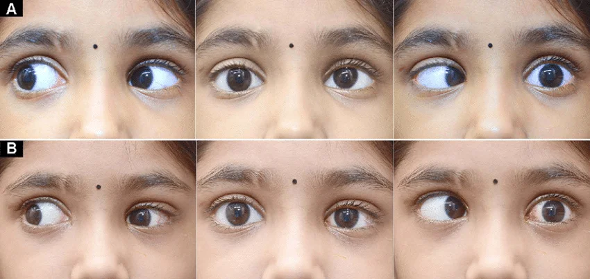
Duane Retraction Syndrome (DRS) Duane Retraction Syndrome (DRS) is a rare genetic condition that affects the muscles that control eye movement. It is characterized by an abnormal movement of the eye, known as retraction, when the eye is attempting to look outwards. This results in the eye appearing to be pulled inward, or “retracted,” during certain eye movements. Symptoms of DRS can include limited or restricted eye movement, double vision, and the appearance of the eye being pulled inward during certain eye movements. The condition can also affect the alignment of the eyes, leading to a cross-eyed or wall-eyed appearance. The diagnosis of DRS is typically made through a comprehensive eye examination, including a thorough examination of the eye muscles and ocular movements. Other tests such as imaging studies, such as an MRI, can also be used to diagnose DRS. Treatment for DRS can include corrective glasses or contact lenses, as well as surgery to reposition the affected eye muscles. The goal of treatment is to improve the alignment of the eyes and reduce symptoms such as double vision. It’s important to note that DRS is a genetic disorder, which is caused by a mutation in the gene that codes for the muscles that control eye movement. Therefore, it is not possible to prevent the occurrence of DRS. Annual health check-ups can be important for individuals with DRS as they may be at risk for certain eye conditions such as amblyopia (lazy eye) or strabismus (crossed eyes). These check-ups can help to identify any early signs of these conditions and allow for early intervention and treatment. Corporate health & wellness programs can also play an important role in the support and care of employees with DRS by providing access to resources and accommodations that can help them to succeed in the workplace. Regular exercise and healthy eating habits are also beneficial for individuals with DRS. Exercise can help to improve overall health and well-being, while a balanced diet can help to maintain a healthy weight and support overall health. In conclusion, Duane Retraction Syndrome (DRS) is a rare genetic disorder that affects the muscles that control eye movement, resulting in retraction of the eye during certain eye movements, double vision and limited or restricted eye movement. DRS is caused by a genetic mutation, which means it’s not preventable. However, annual health check-ups and corporate health & wellness programs can play an important role in the support and care of individuals with DRS by providing access to resources and accommodations that can help them to succeed in the workplace. Regular exercise and healthy eating habits are also beneficial for individuals with DRS. If a person suspects they have DRS, they should seek medical attention as soon as possible.
Read More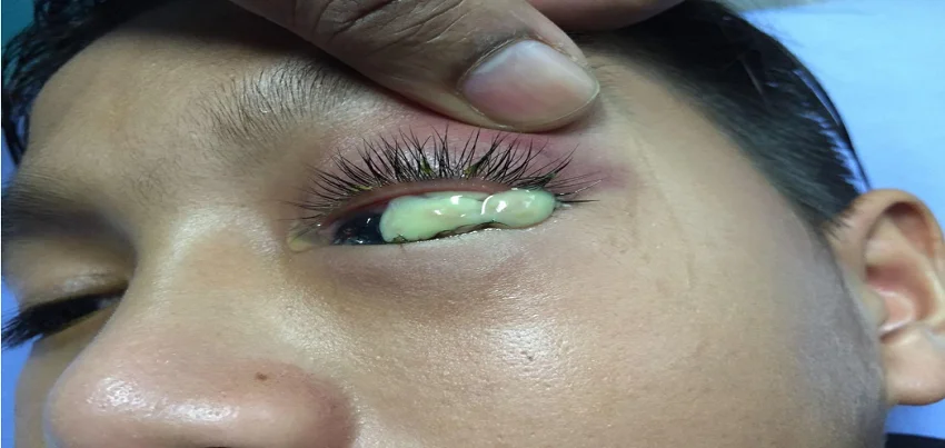
Dacryoadenitis Dacryoadenitis is an inflammation of the lacrimal (tear) gland, which is located in the upper, outer portion of the eye. The condition can cause pain and swelling in the affected area, as well as a reduction in tear production. Symptoms of dacryoadenitis can include pain and swelling in the affected area, redness of the eye, and a reduction in tear production. Other symptoms may include fever, fatigue, and a general feeling of malaise. In some cases, the affected gland may also become infected, leading to the development of an abscess. The diagnosis of dacryoadenitis is typically made through a physical examination and imaging tests such as CT scan or MRI. In some cases, a biopsy may also be performed to confirm the diagnosis. Treatment for dacryoadenitis typically involves the use of antibiotics to clear any infection, as well as anti-inflammatory medications to reduce inflammation and pain. In some cases, surgical intervention may be necessary to drain an abscess or remove the affected gland. It’s important to note that the cause of dacryoadenitis is not well understood, and it is not known how to prevent the condition from occurring. However, maintaining good overall health through healthy lifestyle choices, including a well-balanced diet and regular exercise, may reduce the risk of developing the condition. Annual health check-ups can be important for individuals who have a history of dacryoadenitis or are at risk for developing the condition. These check-ups can help to identify any early signs of the condition and allow for early intervention and treatment. Corporate health & wellness programs can also play an important role in preventing dacryoadenitis by promoting healthy lifestyle choices and providing access to preventive care and health screenings. While there is no specific diet or exercise regimen that has been proven to prevent dacryoadenitis, maintaining a healthy diet and regular exercise can have many benefits for overall health. Eating a diet that is rich in fruits, vegetables, and whole grains can help to support the immune system and reduce the risk of infection. Regular physical activity, such as walking or cycling for 30 minutes a day, can also help to improve overall health and reduce the risk of developing certain conditions. In conclusion, Dacryoadenitis is a condition that causes inflammation of the lacrimal gland, resulting in pain, swelling and tear production reduction. The cause of the condition is not well understood, and it is not known how to prevent it from occurring. However, maintaining good overall health through healthy lifestyle choices, including a well-balanced diet and regular exercise, may reduce the risk of developing the condition. Annual health check-ups and corporate health & wellness programs can play an important role in preventing Dacryoadenitis by promoting healthy lifestyle choices and providing access to preventive care and health screenings. If a person has symptoms, they should seek medical attention as soon as possible.
Read More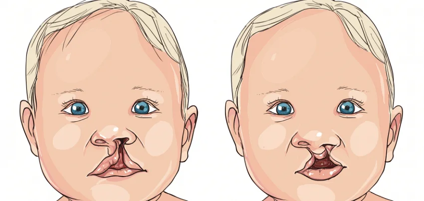
Cleft Lip Cleft lip and cleft palate are congenital conditions that occur when the lip and/or palate do not form properly in the womb, resulting in an opening or “cleft.” Cleft lip and cleft palate can occur separately or together, and can range in severity from a small opening to a complete separation of the lip or palate. Symptoms of cleft lip and cleft palate can vary depending on the severity of the cleft and the specific area affected. Common symptoms include a visible opening or gap in the lip or palate, difficulty with feeding and speech, and ear infections. In some cases, cleft lip and cleft palate may not cause any symptoms at all. Diagnosis of cleft lip and cleft palate is typically made during pregnancy through ultrasound or at birth during physical examination. Prenatal diagnosis can help parents to plan and prepare for the baby’s arrival and the possible need for treatment. Common treatment methods for cleft lip and cleft palate include surgery to repair the cleft, which is typically performed in the first year of life. Additional surgeries may be necessary as the child grows, to address any issues related to speech or breathing. Preventing cleft lip and cleft palate from occurring is not currently possible, as the exact cause of these conditions is not known. However, there are some steps that can be taken to reduce the risk of cleft lip and cleft palate. These include getting early and regular prenatal care, eating a healthy diet during pregnancy, and avoiding smoking and alcohol consumption during pregnancy. Annual health check-ups and corporate health & wellness programs can also play an important role in preventing cleft lip and cleft palate. During these check-ups, a physical examination can be performed to identify any potential risk factors for cleft lip and cleft palate, and make recommendations for prenatal care. In terms of diet and exercise, there is evidence to suggest that a healthy diet during pregnancy, including adequate intake of folic acid and other nutrients, can help to lower the risk of cleft lip and cleft palate. Additionally, regular physical activity can help to improve overall health during pregnancy, which can also have positive effects on the health of the developing baby. It is important to note that cleft lip and cleft palate are conditions that can be treated successfully with surgery, and early intervention can help to improve the outcome for affected individuals. With the help of annual health check-ups and corporate health & wellness programs, early diagnosis and treatment can be ensured for the best possible outcome.
Read More
Congenital Capillary Haemangioma Of The Eyelid Congenital capillary haemangioma of the eyelid, also known as a “strawberry nevus,” is a benign tumor that is present at birth and composed of abnormal blood vessels. It typically appears as a red or purple bump on the eyelid and can range in size from small to large. Symptoms of congenital capillary haemangioma of the eyelid include a visible red or purple bump on the eyelid, as well as a potential loss of eyelashes in the affected area. In rare cases, the tumor may cause vision problems or interfere with the normal functioning of the eye. Diagnosis of congenital capillary haemangioma of the eyelid is typically made through physical examination by an ophthalmologist. In some cases, further testing such as a biopsy may be required to confirm the diagnosis. Common treatment methods for congenital capillary haemangioma of the eyelid include topical and oral medications, laser therapy, and surgery. The most appropriate treatment will depend on the size and location of the tumor, as well as the patient’s overall health. Prevention of congenital capillary haemangioma of the eyelid is not currently possible, as the exact cause of the condition is not known. However, annual health check-ups and corporate health & wellness programs can help identify and treat the condition early on. In terms of diet and exercise, there is currently no evidence to suggest that specific dietary or exercise interventions can prevent the development of congenital capillary haemangioma of the eyelid. However, maintaining a healthy lifestyle through regular physical activity and a balanced diet can have overall benefits for the health of the individual. It’s important to note that congenital capillary haemangioma of the eyelid is a benign condition and treatment is typically successful. However, it is important to seek medical attention if you notice any unusual bumps or changes on or around the eyelid. With the help of annual health check-ups and corporate health & wellness programs, early diagnosis and treatment can be ensured for the best possible outcome.
Read More
Conjunctival Concretions Conjunctival concretions, also known as “conjunctival stones” or “episcleral nodules,” are small, hard lumps that form on the conjunctiva, the clear membrane that covers the white part of the eye and the inside of the eyelids. These concretions are made up of a buildup of protein, calcium, and other substances, and can cause a variety of symptoms, including redness, irritation, and a foreign body sensation in the eye. Symptoms of conjunctival concretions include redness, irritation, and a foreign body sensation in the eye. In some cases, the concretions may cause blurred vision or sensitivity to light. However, some people with conjunctival concretions may not experience any symptoms at all. The diagnosis of conjunctival concretions is typically made by an ophthalmologist during a comprehensive eye exam. The doctor will examine the eye and eyelids for signs of the concretions, and may also use a microscope or other diagnostic tools to get a closer look at the eye. In some cases, a biopsy may be needed to confirm the diagnosis. Common treatment methods for conjunctival concretions include lubricating eye drops, artificial tears, and ointments. These can help to relieve symptoms and keep the eye moist, but they may not be effective in treating the underlying condition. In some cases, surgery may be required to remove the concretions and prevent further complications. To prevent conjunctival concretions from occurring, it is important to maintain good eye health. This includes regular eye exams and annual health check-ups, as well as corporate health and wellness programs that promote healthy living. Diet and exercise also play a role in preventing conjunctival concretions. Eating a diet rich in fruits and vegetables and avoiding foods that are high in fat, sugar, and sodium can help to maintain overall health and reduce the risk of eye problems. Regular exercise, such as brisk walking, cycling, or swimming, can also help to improve blood flow to the eyes and reduce the risk of eye problems. Corporate health and wellness programs can also help to prevent conjunctival concretions by promoting healthy living and encouraging employees to take care of their eyes. These programs may include vision screenings, eye health education, and incentives for regular eye exams and healthy lifestyle choices. In summary, Conjunctival concretions, also known as “conjunctival stones” or “episcleral nodules,” are small, hard lumps that form on the conjunctiva, the clear membrane that covers the white part of the eye and the inside of the eyelids. Symptoms include redness, irritation, and a foreign body sensation in the eye, but some people may not experience any symptoms. The diagnosis of conjunctival concretions is typically made by an ophthalmologist during a comprehensive eye exam, and common treatment methods include lubricating eye drops, artificial tears, and ointments. To prevent conjunctival concretions from occurring, it is important to maintain good eye health, regular eye check-ups, and corporate health and wellness programs that promote healthy living, and a healthy diet and regular exercise. “Understanding Chalcosis: Causes, Symptoms, and Treatment Options Chalcosis, a condition often associated with the presence of metal particles in the eye, can lead to various ocular complications if left untreated. In this article, we’ll explore the causes, symptoms, and treatment options for chalcosis, shedding light on this potentially serious eye condition. Causes of Chalcosis: Chalcosis is primarily caused by the introduction of metallic particles, particularly copper, into the eye. This can occur due to occupational hazards, such as metalworking or welding, where metal fragments or dust may inadvertently come into contact with the eyes. Additionally, chalcosis can result from accidental injury or trauma involving metal objects, leading to the deposition of metal in the cornea or other ocular structures. Symptoms of Chalcosis: The symptoms of chalcosis can vary depending on the severity of the condition and the extent of metal deposition in the eye. Common symptoms may include: Eye irritation and discomfort Redness and inflammation of the affected eye Blurred vision or changes in vision clarity Sensation of a foreign body in the eye Excessive tearing or watery eyes Photophobia (sensitivity to light) In cases where chalcosis progresses, individuals may experience more severe symptoms, such as corneal opacity, cataracts, or other complications affecting vision and ocular health. Diagnosis and Evaluation: Diagnosing chalcosis typically involves a comprehensive eye examination performed by an ophthalmologist. The evaluation may include visual acuity tests, slit-lamp examination, and imaging studies to assess the extent of metal deposition and any associated ocular damage. A thorough medical history, including information about recent eye trauma or exposure to metal particles, is also essential for an accurate diagnosis. Treatment Options for Chalcosis: The treatment approach for chalcosis depends on the severity of the condition and the extent of ocular damage. In mild cases where metal particles are superficially lodged in the eye, treatment may involve: Flushing the eye with saline solution or irrigating solution to remove the metal particles. Topical corticosteroid eye drops to reduce inflammation and promote healing. Antibiotic eye drops or ointments to prevent infection. In more severe cases of chalcosis, where metal particles have penetrated deeper into the cornea or other ocular structures, additional interventions may be required. These may include: Surgical removal of metal fragments or foreign bodies from the eye. Corneal debridement or keratectomy to remove damaged tissue and promote healing. Treatment of associated complications, such as cataracts or glaucoma, if present. Prevention and Eye Safety Measures: Preventing chalcosis involves implementing appropriate eye safety measures, especially in occupational settings where exposure to metal particles is common. This includes wearing protective eyewear, such as safety goggles or face shields, to prevent foreign bodies from entering the eyes during metalworking or other high-risk activities. Regular eye examinations and prompt medical attention in the event of eye trauma or injury are also crucial for early detection and treatment of chalcosis. In conclusion, chalcosis is a potentially serious eye condition caused by the deposition of metal particles in the eye. Understanding the causes, symptoms, and treatment options for chalcosis is essential for timely intervention and preservation of ocular health.
Read More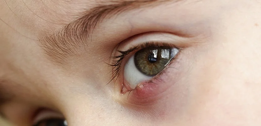
Chalazion Chalazion, also known as a meibomian cyst, is a small, painless lump that develops on the eyelid as a result of a blocked oil gland. They are relatively common and can occur on both the upper and lower eyelids. Chalazions can cause swelling and redness of the eyelid, and can also cause vision problems if they grow large enough to press on the eye. Symptoms of a chalazion include a small, painless lump on the eyelid, swelling and redness of the eyelid, and in some cases, vision problems. The lump may be tender to the touch, and may cause the eyelid to droop. Diagnosis of a chalazion is typically made through a physical examination of the eyelid. In some cases, a biopsy may be performed to rule out other conditions such as skin cancer. Treatment for a chalazion typically involves the application of warm compresses to the affected area, which can help to soften and open the blocked gland. In some cases, an antibiotic ointment may be prescribed. If the chalazion does not respond to these treatments, surgery may be performed to remove the cyst. To prevent chalazion from occurring, it is important to maintain good eyelid hygiene, which includes regularly cleaning the eyelids with a mild soap and water and avoiding the use of eye makeup. Annual health check-ups can also help to identify any underlying conditions that may increase the risk of chalazion, such as blepharitis (eyelid inflammation) and rosacea. Corporate health and wellness programs can also help to promote healthy habits and reduce the risk of eyelid problems. Diet and exercise can also play a role in preventing chalazion. A healthy diet that is rich in fruits, vegetables, and omega-3 fatty acids can help to promote good eye health and reduce the risk of eyelid problems. Regular exercise can also help to improve overall health and reduce the risk of chronic conditions such as obesity, which can increase the risk of chalazion. In summary, Chalazion is a small, painless lump that develops on the eyelid as a result of a blocked oil gland. They are relatively common and can occur on both the upper and lower eyelids. Symptoms include a small, painless lump on the eyelid, swelling and redness of the eyelid, and in some cases, vision problems. Diagnosis is typically made through a physical examination of the eyelid. Treatment usually involves application of warm compresses to the affected area, antibiotics ointment and in some cases surgery. To prevent chalazion from occurring, maintain good eyelid hygiene, annual health check-ups, corporate health and wellness programs and a healthy diet rich in fruits, vegetables, and omega-3 fatty acids can help to promote good eye health and reduce the risk of eyelid problems. Regular exercise also helps to improve overall health and reduce the risk of chronic conditions such as obesity which can increase the risk of chalazion.
Read More
Bedsores Bedsores, also known as pressure ulcers, are injuries to the skin and underlying tissue that occur when prolonged pressure is applied to an area of the body. They are most commonly found on bony areas of the body such as the hips, heels, and tailbone and are most common in individuals who are bedridden or have limited mobility. In this article, we will discuss the symptoms of bedsores, tests for diagnosis, common treatment methods, and ways to prevent them from occurring. Additionally, we will discuss the importance of diet and exercise in maintaining overall health through corporate health and wellness programs. Symptoms of bedsores can vary depending on the stage of the injury, but common symptoms include redness, warmth, and pain in the affected area. As the injury progresses, the skin may become broken, and an open wound may develop. In advanced stages, the wound may be deep and expose underlying tissue. Diagnosis of bedsores is typically made through visual examination of the affected area. In some cases, additional tests such as a wound culture or tissue biopsy may be performed to identify any underlying infections or complications. Common treatment methods for bedsores include cleaning and dressing the wound, preventing further pressure on the affected area, and managing any underlying infections. In some cases, surgery may be necessary to remove dead tissue or to close the wound. Prevention of bedsores is crucial, and it can be done through frequent repositioning of the individual, maintaining good hygiene and proper skin care, and providing adequate nutrition and hydration. Corporate health and wellness programs can play a role in preventing bedsores by providing education on the importance of proper skin care and positioning, and offering support and resources to assist individuals with limited mobility. Diet and exercise are also important in maintaining overall health and reducing the risk of bedsores. Eating a balanced diet that includes plenty of fruits and vegetables, lean protein, and whole grains can help to support the skin and immune system. Regular exercise can also help to improve overall health and reduce the risk of chronic disease. In conclusion, bedsores are a serious condition that can cause significant discomfort and lead to serious complications if not properly treated. Through preventative measures such as maintaining proper positioning, proper hygiene, and good nutrition, and participating in corporate health and wellness programs, individuals can take steps to reduce their risk of developing bedsores. If you suspect that you or a loved one is at risk for bedsores, it is important to seek medical attention as soon as possible for proper diagnosis and treatment.
Read More
Acquired Capillary Haemangioma Of The Eyelid Acquired capillary haemangioma of the eyelid, also known as a “strawberry hemangioma,” is a benign (non-cancerous) growth that occurs on the skin of the eyelid. These growths are made up of small, dilated blood vessels and can vary in size and color. They are more common in infants and young children, but can occur in adults as well. Symptoms of a strawberry hemangioma include a raised, red or purple bump on the eyelid. In some cases, the growth may be itchy or cause vision problems. Diagnosis of a strawberry hemangioma is typically made by a physical examination and may be confirmed with a biopsy. Common treatment methods for strawberry hemangiomas include topical or oral steroids, laser therapy, and surgery. The choice of treatment will depend on the size, location, and symptoms of the growth. Prevention of strawberry hemangiomas is not possible, as the cause is not well understood. However, regular skin exams and health check-ups can help to detect and diagnose the growth early. Diet and exercise have not been shown to prevent the development of strawberry hemangiomas. However, a healthy diet and regular exercise can help to maintain overall health and well-being. Incorporating regular skin exams and health check-ups into corporate health and wellness programs can help to detect and diagnose strawberry hemangiomas early, allowing for prompt treatment and management. This can help to minimize the impact of the growth on an individual’s quality of life and prevent vision problems from occurring.
Read MoreTop rated products
-
 Full Body Health Checkup I
Original price was: ₹3,000.00.₹1,770.00Current price is: ₹1,770.00.
Full Body Health Checkup I
Original price was: ₹3,000.00.₹1,770.00Current price is: ₹1,770.00.
-
Full Body Health Checkup V
₹3,900.00
-
Pro Health Plus Female
Original price was: ₹15,000.00.₹8,250.00Current price is: ₹8,250.00.
-
Healthy life Plus Male
Original price was: ₹15,000.00.₹8,250.00Current price is: ₹8,250.00.
-
Progno Health AHC I Annual
Original price was: ₹3,000.00.₹1,650.00Current price is: ₹1,650.00.


-
₹999.00
-
₹16,000.00Original price was: ₹16,000.00.₹5,910.00Current price is: ₹5,910.00. -
₹849.00
-
₹1,999.00
-
₹2,999.00