PrognoHealth – Corporate Health & Wellness Specialist

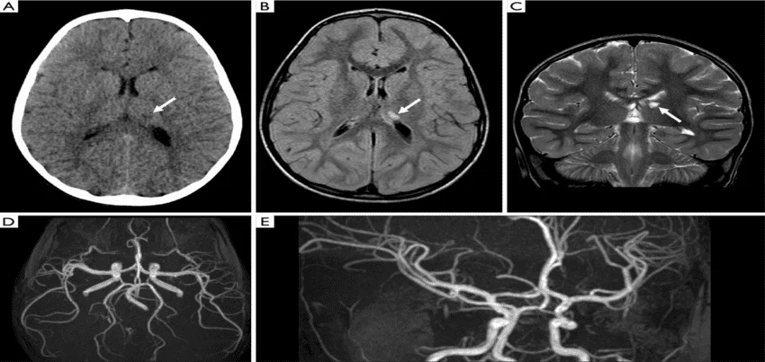
MRI Angiography Of Brain Or MRA Of Brain Magnetic Resonance Angiography (MRA) of the brain is a non-invasive medical imaging test that uses a powerful magnetic field, radio waves, and a computer to create detailed images of the blood vessels in the brain. It provides valuable information about the structure and function of the blood vessels, helping physicians diagnose and treat a variety of conditions. The Test Procedure: During an MRA of the brain, the patient lies on a table that slides into a tunnel-shaped machine. The patient’s head is secured with straps, and they are provided with earplugs to protect against the loud banging and knocking noises the machine makes during the procedure. The test typically takes between 30 to 60 minutes to complete, depending on the complexity of the images required. To create clear and detailed images, the patient must remain still during the test. The technician will provide the patient with instructions and may use a communication system to keep in touch with the patient during the procedure. Test Preparation: Preparation for an MRA of the brain may vary depending on the type of scan ordered by the physician. Generally, the patient is instructed to avoid wearing any metallic objects, such as jewelry, watches, or clothing with metal zippers, during the procedure. Patients should also inform the technician if they have any implanted medical devices, such as pacemakers, as these may interfere with the MRI machine. Patients may be asked to avoid eating or drinking for several hours before the test, especially if they are undergoing a contrast-enhanced scan. The technician will provide specific instructions to the patient before the test. Common Symptoms for Ordering the Test: An MRA of the brain may be ordered by a physician for a variety of reasons, including the following symptoms: HeadachesDizziness or vertigoNumbness or tinglingVision problemsMemory loss or confusionSuspected aneurysm or other vascular abnormalitiesSuspected stroke or other cerebrovascular disordersSuspected arteriovenous malformations (AVMs) or other vascular tumors Health Checkup, Wellness, and Corporate Health: An MRA of the brain may be included as part of a comprehensive health checkup or wellness program, especially for individuals at higher risk of cerebrovascular disorders. Corporate health programs may also offer MRA of the brain as a screening tool for employees, especially those who work in high-stress or high-risk environments. The results of an MRA of the brain may help physicians identify potential health risks and develop personalized treatment plans. In some cases, early detection of cerebrovascular disorders can improve the effectiveness of treatment and increase the chances of successful recovery. Interpretation of Results: Interpreting the results of an MRA of the brain requires specialized training and expertise. The images produced by the MRA machine are highly detailed and may reveal subtle changes in the blood vessels of the brain. A radiologist or neurologist typically reviews the images and provides a report to the referring physician. The results of an MRA of the brain may indicate the presence of aneurysms, AVMs, or other vascular abnormalities that require further testing or treatment. The images may also reveal evidence of stroke or other cerebrovascular disorders, such as narrowing or blockage of the blood vessels in the brain. In some cases, the results of an MRA of the brain may be inconclusive or require further testing or evaluation. The physician will discuss the results with the patient and develop a treatment plan based on the findings. In conclusion, an MRA of the brain is a valuable diagnostic tool that provides detailed information about the blood vessels in the brain. The test is non-invasive and painless, making it an ideal screening tool for individuals “Demystifying MRA vs. MRI: Understanding the Differences and Applications In the realm of diagnostic imaging, Magnetic Resonance Imaging (MRI) and Magnetic Resonance Angiography (MRA) stand out as crucial tools for understanding the intricacies of the human body, especially the brain’s vascular system. While they both utilize magnetic fields and radio waves to generate detailed images, they serve distinct purposes. In this blog, we’ll delve into the disparities between MRI and MRA, explore their individual applications, and discuss how they complement each other in diagnosing various neurovascular conditions. Understanding MRI and MRA: Magnetic Resonance Imaging (MRI): MRI is a versatile imaging technique used to visualize detailed internal structures of the body. It provides high-resolution images of soft tissues, organs, bones, and even the brain without using ionizing radiation. MRI works by aligning the body’s hydrogen atoms with a strong magnetic field and then applying radio waves to create signals that are processed into detailed images by a computer. Magnetic Resonance Angiography (MRA): On the other hand, MRA is a specialized form of MRI primarily focused on imaging blood vessels. It provides detailed images of blood flow within arteries and veins, offering insights into the vascular anatomy without invasive procedures. MRA employs techniques such as time-of-flight, contrast-enhanced, or phase-contrast imaging to visualize blood vessels and detect abnormalities. Differences between MRI and MRA: Purpose: MRI is used to examine a wide range of anatomical structures and soft tissues. MRA specifically targets blood vessels, providing detailed images of the vascular system’s anatomy and blood flow. Contrast Agents: While MRI can use contrast agents to enhance image quality for certain examinations, it’s not always necessary. MRA often utilizes contrast agents to highlight blood vessels and detect abnormalities more effectively. Techniques: MRI employs various techniques such as T1-weighted, T2-weighted, and diffusion-weighted imaging to visualize different tissue characteristics. MRA utilizes specific techniques like time-of-flight or contrast-enhanced imaging to highlight blood vessels’ contrast with surrounding tissues. Applications: MRI is widely used for diagnosing conditions ranging from musculoskeletal injuries to neurological disorders. MRA is particularly valuable for diagnosing vascular conditions like aneurysms, stenosis, or arteriovenous malformations (AVMs). Can MRI and MRA be done together? Yes, MRI and MRA can be performed sequentially during the same imaging session. This allows comprehensive evaluation of both anatomical structures and vascular flow patterns without requiring separate appointments, providing a holistic assessment for patients with suspected neurovascular disorders. Clinical
Read More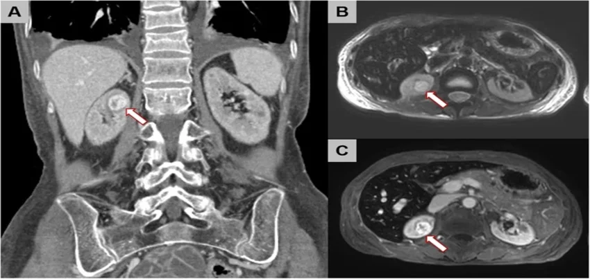
MRI Of The Abdomen Magnetic Resonance Imaging (MRI) is a non-invasive medical imaging technique that uses a strong magnetic field and radio waves to produce detailed images of the inside of the body. An MRI of the abdomen is a diagnostic test that uses this technology to capture images of the organs and tissues in the abdominal region. The test is typically conducted in a hospital or diagnostic imaging center. Before the procedure, patients are asked to remove any metal objects, such as jewelry, watches, and hairpins, as these can interfere with the magnetic field of the machine. Patients may also be asked to change into a hospital gown or other loose-fitting clothing. During the test, the patient lies down on a narrow table that slides into the MRI machine. The machine creates a strong magnetic field around the body, which causes the protons in the body’s tissues to align themselves. The radio waves are then used to stimulate the protons, causing them to emit a signal that is picked up by the machine’s sensors. This signal is used to create detailed images of the inside of the body, which can be viewed by a radiologist. In preparation for an MRI of the abdomen, patients may be asked to fast for a period of time before the procedure. This is to ensure that the stomach and intestines are empty, which can improve the clarity of the images. Patients may also be asked to drink a special dye, known as contrast material, which can help to highlight certain areas of the abdomen and improve the accuracy of the images. The most common symptoms that may lead to an MRI of the abdomen include abdominal pain, bloating, diarrhea, constipation, and unexplained weight loss. These symptoms may be indicative of a variety of conditions, such as inflammatory bowel disease, liver disease, or tumors. The length of time that an MRI of the abdomen takes can vary depending on a number of factors, such as the size of the area being imaged and the quality of the images needed. Typically, the test can take between 30 minutes and an hour to complete. After the test is complete, a radiologist will review the images and provide a report to the patient’s doctor. The report will include information about any abnormalities that were found, as well as recommendations for further testing or treatment if necessary. An MRI of the abdomen can be an important part of a health checkup, wellness program, or corporate health initiative. By identifying potential health issues early on, patients can take steps to address them and improve their overall health and wellbeing. Additionally, regular MRI screenings may be recommended for patients with a family history of certain conditions or who are at a higher risk of developing certain health problems. In conclusion, an MRI of the abdomen is a non-invasive diagnostic test that uses magnetic fields and radio waves to produce detailed images of the organs and tissues in the abdominal region. The test can be conducted in a hospital or imaging center, and patients may be asked to fast or drink a special dye before the procedure. Common symptoms that may lead to an MRI of the abdomen include abdominal pain, bloating, and unexplained weight loss. The length of the test can vary, and results are typically provided to the patient’s doctor for further evaluation and treatment. By incorporating regular MRI screenings into health checkups, wellness programs, and corporate health initiatives, patients can take proactive steps to maintain their health and wellbeing.
Read More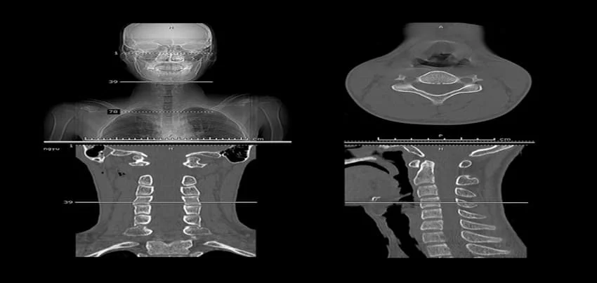
CT Scan Of The Neck A CT (computed tomography) scan of the neck is a diagnostic imaging test that uses X-rays and computer technology to produce detailed images of the neck area. This test is commonly ordered when a patient is experiencing symptoms related to the neck or throat. Test Conducted and Preparation: During a CT scan of the neck, the patient lies down on a table that slides into the CT machine. The machine takes multiple images from different angles and uses computer processing to create a 3D image of the neck. The test is painless and usually takes less than 30 minutes to complete. There is no specific preparation required for a CT scan of the neck. However, patients may be asked to remove any metal objects, such as jewelry or eyeglasses, before the test. Common Symptoms for Ordering the Test: A CT scan of the neck may be ordered by a doctor if a patient is experiencing symptoms such as: Neck pain or stiffnessSwelling or lumps in the neckDifficulty swallowingHoarseness or voice changesUnexplained weight lossHistory of smoking or alcohol abuseTrauma to the neckThese symptoms could indicate conditions such as tumors, infections, or damage to the neck area. Time Taken for the Test and its Results Interpretations: The test itself takes only a few minutes, but the entire process, including check-in, preparation, and post-test consultation, may take up to an hour. After the test, a radiologist will review the images and prepare a report for the patient’s doctor. The doctor will then discuss the results with the patient and provide any necessary treatment recommendations. Using Health Checkup, Wellness, and Corporate Health as Key Words: A CT scan of the neck may be included as part of a comprehensive health checkup or wellness program. It can help detect and diagnose conditions such as tumors, infections, and abnormalities in the neck area. For corporate health programs, this test may be used to assess employees’ health risks and provide preventive care. In addition, a CT scan of the neck can be useful for monitoring the effectiveness of treatments for conditions such as cancer or infections. It can also be used to guide surgical procedures or biopsies of the neck area. While a CT scan of the neck is generally considered a safe and effective diagnostic tool, it does involve exposure to ionizing radiation. Therefore, doctors typically limit the number of CT scans a patient receives to reduce the risk of radiation-related side effects. Patients should always discuss the risks and benefits of any imaging test with their doctor. In conclusion, a CT scan of the neck is a valuable diagnostic tool that can provide detailed images of the neck area. It is a quick and painless test that can help diagnose a range of conditions related to the neck or throat. This test may be included as part of a health checkup or wellness program and can be useful in corporate health settings to assess and manage employee health risks. It is important for patients to discuss the risks and benefits of any imaging test with their doctor and to follow any preparation instructions provided prior to the test.
Read More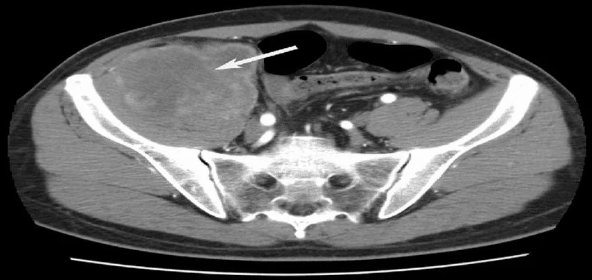
CT Scan of the Pelvis A CT scan of the pelvis is a diagnostic imaging test that uses X-rays and computer technology to create detailed images of the pelvic area. This test is commonly ordered when a patient is experiencing symptoms related to the pelvic region or when a doctor wants to investigate a potential health issue in this area. Test Conducted and Preparation: During a CT scan of the pelvis, the patient lies down on a table that slides into the CT machine. The machine takes multiple images from different angles and uses computer processing to create a 3D image of the pelvic area. The test is painless and usually takes less than 30 minutes to complete. In some cases, the doctor may order a contrast dye to be injected into the patient’s vein prior to the test to help highlight certain structures in the pelvic area. In these cases, the patient may be asked to fast for a few hours before the test and to drink plenty of fluids to help flush the dye from their system after the test. Common Symptoms for Ordering the Test: A CT scan of the pelvis may be ordered by a doctor if a patient is experiencing symptoms such as: Pain or discomfort in the pelvic areaAbnormal vaginal bleedingDifficulty urinatingUnexplained weight lossSwelling or lumps in the pelvic areaThese symptoms could indicate conditions such as infections, tumors, or injuries to the pelvic area. Time Taken for the Test and its Results Interpretations: The test itself takes only a few minutes, but the entire process, including check-in, preparation, and post-test consultation, may take up to an hour. After the test, a radiologist will review the images and prepare a report for the patient’s doctor. The doctor will then discuss the results with the patient and provide any necessary treatment recommendations. Using Health Checkup, Wellness, and Corporate Health as Key Words: A CT scan of the pelvis may be included as part of a comprehensive health checkup or wellness program. It can help detect and diagnose conditions such as infections, tumors, and injuries to the pelvic area. For corporate health programs, this test may be used to assess employees’ health risks and provide preventive care. In addition, a CT scan of the pelvis can be useful for monitoring the effectiveness of treatments for conditions such as tumors or infections. It can also be used to guide surgical procedures or biopsies of the pelvic area. While a CT scan of the pelvis is generally considered a safe and effective diagnostic tool, it does involve exposure to ionizing radiation. Therefore, doctors typically limit the number of CT scans a patient receives to reduce the risk of radiation-related side effects. Patients should always discuss the risks and benefits of any imaging test with their doctor. In conclusion, a CT scan of the pelvis is a valuable diagnostic tool that can provide detailed images of the pelvic area. It is a quick and painless test that can help diagnose a range of conditions related to the pelvic region. This test may be included as part of a health checkup or wellness program and can be useful in corporate health settings to assess and manage employee health risks. It is important for patients to discuss the risks and benefits of any imaging test with their doctor and to follow any preparation instructions provided prior to the test.
Read More
Tuberculosis Tuberculosis, also known as TB, is a bacterial infection that primarily affects the lungs. However, it can also affect other parts of the body such as the kidneys, spine, and brain. It is a serious and potentially deadly disease, but it is also preventable and treatable. Symptoms of TB include a persistent cough that lasts for more than three weeks, chest pain, weight loss, fatigue, night sweats, and fever. If you experience any of these symptoms, it is important to seek medical attention as soon as possible. Diagnosis of TB is typically done through a combination of tests, including a chest X-ray, a skin test called the tuberculin test, and a blood test called the interferon-gamma release assay. Sputum culture may also be performed to identify the specific strain of bacteria causing the infection. Common treatment methods for TB include a combination of antibiotics, which are typically taken for six to nine months. It is important to complete the full course of antibiotics even if you start feeling better, as this can help prevent the development of drug-resistant TB. Preventing TB from occurring is possible through a combination of vaccination, healthy lifestyle choices, and early detection and treatment of the disease. The Bacillus Calmette-Guérin (BCG) vaccine is given to children in many countries to protect against TB, and it is also recommended for adults at high risk of the disease. To prevent TB, it’s important to maintain a healthy diet, exercise regularly, and avoid smoking and excessive alcohol consumption. Annual health check-ups and corporate health & wellness programs can also help identify and prevent TB. Diet and exercise can also play a role in preventing TB. Eating a diet rich in fruits and vegetables, lean protein, and whole grains can help boost your immune system and protect against infection. Regular exercise can also help keep your lungs healthy and reduce your risk of developing TB. In conclusion, tuberculosis is a serious and potentially deadly disease, but it is also preventable and treatable. Regular check-ups, healthy lifestyle choices, and early detection and treatment are key to preventing TB. Corporate health and wellness programs can also play an important role in identifying and preventing TB among employees.
Read More
Lipoprotein (a) Test Lipoprotein (a), or Lp(a), is a type of low-density lipoprotein (LDL) that is associated with an increased risk of heart disease. The Lipoprotein (a) test is used to measure the level of Lp(a) in the blood. Pre-test preparation: There is no specific pre-test preparation required for the Lipoprotein (a) test. The test is typically done by drawing blood from a vein in the arm and sending it to a lab for analysis. Common symptoms: That may lead a healthcare provider to prescribe Lipoprotein (a) test include high blood pressure, high cholesterol, diabetes, smoking, obesity, family history of heart disease, and age above 45 years old. Diagnosis: The test is used to identify people who have high levels of Lp(a) and are at an increased risk of heart disease. High levels of Lp(a) can be inherited and can’t be reduced with lifestyle changes or medication. However, a healthcare provider can recommend preventive measures to reduce the risk of heart disease. Reference range: The reference range for Lp(a) varies depending on the lab that is performing the analysis. However, a normal value is considered less than 30 mg/dL. Values above that level are considered high and may indicate an increased risk of heart disease. Medical disclaimer: It’s important to note that Lp(a) is only one of many risk factors for heart disease, so a healthcare professional will consider other risk factors as well. Additionally, other medical conditions can also affect the results of Lp(a) test, so it is important to consult a healthcare professional for proper interpretation of the results.
Read More
Lead Level Test A Lead Level test is a blood test that measures the amount of lead in the blood. Lead is a toxic heavy metal that can have serious health effects, particularly in children and pregnant women. Pre-test preparation: There is no specific preparation required for a lead level test. Testing method: A small sample of blood is taken from a vein in your arm and sent to a laboratory for analysis. Common symptoms: That may prompt your doctor to order a lead level test include: abdominal pain, constipation, headache, irritability, and fatigue. Diagnosis: The lead level test is used to screen for and diagnose lead poisoning, which occurs when lead builds up in the body over time. It is also used to monitor treatment for lead poisoning. Reference range: The normal range for lead levels in the blood is considered to be less than 5 micrograms per deciliter (µg/dL). However, it is worth noting that no level of lead exposure is considered safe, and even low levels of lead can have health effects. Normal values may vary depending on the laboratory that performs the test. Medical disclaimer: The information provided is not intended to be a substitute for professional medical advice, diagnosis, or treatment. Always seek the advice of your physician or other qualified healthcare provider with any questions you may have regarding a medical condition. It is important to follow the instructions given by your doctor or the laboratory performing the test. If you have symptoms that suggest lead poisoning or have been advised to have a lead level test, it is important to work closely with your doctor to understand the results.
Read More
Troponin T Test Troponin T is a protein found in heart muscle cells. A Troponin T test is a blood test that measures the amount of Troponin T in the blood. This test is often used to help diagnose a heart attack or to assess the extent of heart damage after a heart attack. Pre-test preparation: Typically does not require any special preparation. The test is performed on a blood sample taken from the patient. Testing method: Typically involves measuring the amount of Troponin T in the blood using a sensitive assay. The test can be performed on a small blood sample, and results are usually available within a few hours. Common symptoms that may prompt a healthcare provider to prescribe a Troponin T test: Include chest pain or discomfort, shortness of breath, or fainting. Other symptoms may include nausea, vomiting, or arm or jaw pain. Diagnosis: Elevated levels of Troponin T in the blood can indicate damage to the heart muscle, which is usually caused by a heart attack. However, other conditions such as severe heart failure, kidney disease, or sepsis can also cause elevated Troponin T levels. Reference range: The reference range for Troponin T levels in the blood is typically less than 0.01 ng/mL. However, it’s important to note that this can vary based on the lab and the specific test used. Normal values: A normal value for Troponin T is considered to be less than 0.01 ng/mL. However, it’s important to note that even small elevations in Troponin T levels can indicate heart damage and should be evaluated by a healthcare professional. Medical disclaimer: It’s also important to consult with a healthcare professional to interpret test results and to understand how they may be affected by individual factors such as medical history and other lab test results.
Read More
Endometriosis Endometriosis is a condition in which the tissue that lines the uterus, called the endometrium, grows outside of the uterus. This tissue can be found on the ovaries, fallopian tubes, and even in the abdominal cavity. Endometriosis can cause a variety of symptoms, including pelvic pain, heavy menstrual bleeding, and infertility. Symptoms of endometriosis include: Pelvic pain and cramping that can occur before, during, or after menstruation Heavy menstrual bleeding Infertility Pain during intercourse Painful bowel movements or urination during menstruation Fatigue and/or depression Endometriosis can be difficult to diagnose, as symptoms can be similar to those of other conditions. A pelvic exam, ultrasound, and/or MRI can be used to help diagnose the condition. In some cases, a laparoscopy, a surgical procedure in which a small camera is inserted into the abdomen, may be necessary to confirm the diagnosis. Common treatment methods for endometriosis include: Hormonal therapy, which can help to reduce the growth of endometrial tissue and alleviate pain Pain medication to help manage pain and cramping Surgery to remove endometrial tissue, which may be necessary in severe cases Preventing endometriosis from occurring is difficult, as the exact cause of the condition is not known. However, there are steps that can be taken to reduce the risk of developing the condition. These include:Annual health check-ups, as early detection and treatment can improve outcomes Corporate health and wellness programs, which can help employees to maintain a healthy lifestyle and reduce the risk of developing chronic health conditions Diet and exercise can also play a role in preventing endometriosis. A healthy diet that is rich in fruits, vegetables, and whole grains can help to reduce inflammation and promote overall health. Regular exercise can also help to reduce the risk of developing endometriosis, as well as alleviate symptoms of the condition. Overall, endometriosis is a complex condition that can cause a variety of symptoms. It is often difficult to diagnose and can be challenging to treat. However, annual health check-ups and corporate health and wellness programs can play a key role in detecting the condition early and reducing the risk of developing endometriosis. Additionally, a healthy diet and regular exercise can help to prevent the condition and alleviate symptoms.
Read More
Bone Fracture A bone fracture, or simply a fracture, is a medical condition in which a bone is broken or cracked. The symptoms of a fracture can vary depending on the location and severity of the injury, but common symptoms include pain, swelling, bruising, and difficulty using the affected limb. Diagnosis of a fracture typically involves a physical examination and imaging tests such as X-rays, CT scans, and MRIs. These tests can help to determine the location and severity of the fracture, as well as any associated injuries or complications. Treatment for a fracture typically involves immobilization of the affected limb, either through the use of a cast or brace, or through surgery. Pain medication may also be prescribed to help manage discomfort. In some cases, physical therapy may be recommended to help restore function and strength to the affected limb. To prevent fractures from occurring, it is important to maintain a healthy diet and exercise regularly. This includes getting enough calcium and vitamin D to support bone health, as well as engaging in weight-bearing exercises to strengthen bones and improve balance. Annual health check-ups can also be helpful in preventing fractures by identifying potential risk factors and providing guidance on how to address them. This can include screenings for conditions such as osteoporosis, which can increase the risk of fractures. Corporate health and wellness programs can also play a role in preventing fractures by promoting healthy behaviors and providing resources for employees to improve their overall health and well-being. This can include offering incentives for regular exercise, providing education on healthy eating, and offering on-site health screenings and assessments. In addition to diet and exercise, there are other lifestyle changes that can help to prevent fractures. Quitting smoking, limiting alcohol consumption, and maintaining a healthy weight can all help to reduce the risk of fractures. When it comes to diet, it is important to get enough calcium and vitamin D. Calcium is important for bone health, and vitamin D helps the body to absorb calcium. Good sources of calcium include dairy products, leafy green vegetables, and fish with edible bones (such as canned salmon or sardines). Good sources of vitamin D include fatty fish, egg yolks, and fortified foods (such as some brands of milk, orange juice, and cereal). Exercise is also important for preventing fractures. Weight-bearing exercises (such as walking, jogging, or dancing) can help to improve bone density and reduce the risk of fractures. Additionally, exercises that improve balance (such as tai chi or yoga) can help to reduce the risk of falls, which can lead to fractures. By incorporating healthy diet, regular exercise and annual health check-ups, as well as corporate health and wellness programs, individuals can take steps to reduce their risk of fractures and improve their overall health and well-being.
Read MoreTop rated products
-
 Full Body Health Checkup I
Original price was: ₹3,000.00.₹1,770.00Current price is: ₹1,770.00.
Full Body Health Checkup I
Original price was: ₹3,000.00.₹1,770.00Current price is: ₹1,770.00.
-
 Young Life Basic
Original price was: ₹3,000.00.₹1,350.00Current price is: ₹1,350.00.
Young Life Basic
Original price was: ₹3,000.00.₹1,350.00Current price is: ₹1,350.00.
-
 COVID 19 Rapid Antigen Test
Original price was: ₹668.00.₹501.00Current price is: ₹501.00.
COVID 19 Rapid Antigen Test
Original price was: ₹668.00.₹501.00Current price is: ₹501.00.
-
 MFG IND PACK III
Original price was: ₹3,900.00.₹2,145.00Current price is: ₹2,145.00.
MFG IND PACK III
Original price was: ₹3,900.00.₹2,145.00Current price is: ₹2,145.00.
-
 Healthy Life Advance Male
Original price was: ₹15,000.00.₹8,250.00Current price is: ₹8,250.00.
Healthy Life Advance Male
Original price was: ₹15,000.00.₹8,250.00Current price is: ₹8,250.00.