PrognoHealth – Corporate Health & Wellness Specialist


CT scan of the abdomen with contrast A CT scan of the abdomen with contrast is a specialized medical imaging test that is used to evaluate the organs and structures within the abdomen. This test combines X-ray technology with the use of a contrast dye, which helps to enhance the images of the organs and structures. In this blog post, we will discuss how this test is conducted, what are the common symptoms for ordering the test, how to prepare for the test, how long does it take, and how the results are interpreted. Test procedure and preparation : Before the CT scan, the patient may be asked to remove any metal objects, such as jewelry or dentures, and to wear a hospital gown. The patient will then lie down on a table that slides into a large, doughnut-shaped machine that emits X-rays. During the scan, the patient may be asked to hold their breath or to stay still to avoid any blurring of the images. In some cases, the doctor may require the use of contrast dye, which helps to highlight the blood vessels and organs within the abdomen. The contrast dye may be given orally or intravenously, depending on the reason for the test. The patient may also be asked to fast for several hours before the test to avoid any interference with the absorption of the contrast dye. Common symptoms for ordering the test : A CT scan of the abdomen with contrast may be ordered by a doctor if the patient is experiencing symptoms such as abdominal pain, bloating, nausea, vomiting, fever, or unexplained weight loss. This test may also be used to monitor the progression of a known condition or to evaluate the effectiveness of a treatment. Time taken for the test and results interpretation : The duration of the CT scan of the abdomen with contrast depends on the complexity of the images required and whether or not a contrast dye is used. Typically, the test takes between 30 minutes to an hour to complete. After the test, the images will be reviewed by a radiologist, who will provide a report to the doctor. The doctor will then discuss the results with the patient, which may include further testing, treatment, or referral to a specialist. Using health checkup, wellness, and corporate health as keywords : A CT scan of the abdomen with contrast may be included in a comprehensive health checkup, which is a series of medical tests and screenings that are performed to evaluate a person’s overall health and well-being. A health checkup may also include blood tests, urine tests, electrocardiograms, and other imaging tests, such as a mammogram or a bone density scan. In the context of corporate health and wellness, a CT scan of the abdomen with contrast may be offered as part of an employee health program, which aims to promote the health and productivity of the workforce. This program may include regular health screenings, wellness coaching, fitness classes, and other health-related services. In conclusion, a CT scan of the abdomen with contrast is a valuable diagnostic tool that can help doctors diagnose and monitor a variety of conditions within the abdomen. If you are experiencing any symptoms that may indicate a problem in the abdomen, or if you are due for a routine health checkup, speak with your doctor about whether a CT scan may be appropriate for you “Understanding Abdominal CT Scans with Contrast: Procedure, Benefits, and Interpretation Introduction: Abdominal CT scans with contrast play a crucial role in diagnosing and evaluating a wide range of abdominal conditions. This comprehensive guide provides insights into the procedure, benefits, and interpretation of abdominal CT scans with contrast, shedding light on their importance in clinical practice. What is Contrast in CT Scan? Contrast in CT scans refers to a substance, typically iodine-based, that is administered intravenously to enhance the visibility of blood vessels, organs, and tissues on imaging. Contrast helps differentiate between normal and abnormal structures, improving the accuracy of diagnosis and assessment. Procedure for Abdominal CT Scan with Contrast: Before the scan, patients may be asked to fast for a few hours to ensure optimal imaging quality. Once in the CT scanner, a contrast agent is injected intravenously, followed by sequential imaging of the abdomen. The contrast material highlights blood vessels and enhances the visualization of organs and tissues, providing detailed images for interpretation by radiologists. Benefits of Abdominal CT Scan with Contrast: Abdominal CT scans with contrast offer several benefits: Improved Visualization: Contrast enhances the visibility of blood vessels, organs, and abnormalities, allowing for more accurate diagnosis and assessment. Enhanced Diagnostic Accuracy: The use of contrast helps differentiate between normal and abnormal structures, aiding in the detection and characterization of various conditions. Comprehensive Evaluation: Contrast-enhanced CT scans provide detailed images of the abdomen, facilitating the assessment of tumors, inflammation, infection, and other pathologies. Treatment Planning: The detailed images obtained from contrast-enhanced CT scans help guide treatment planning and interventions, such as surgery, chemotherapy, or radiation therapy. Monitoring Response to Treatment: Follow-up CT scans with contrast can monitor the response to treatment and detect any changes in the size or characteristics of abnormalities over time. Preparation for Abdominal CT Scan with Contrast: Patients may be instructed to fast for several hours before the scan and avoid certain medications that could interfere with the imaging process. It’s essential to inform the healthcare team about any allergies, pre-existing medical conditions, or medications being taken, as well as any previous adverse reactions to contrast agents. Interpretation of Abdominal CT Scan with Contrast: Radiologists interpret the images obtained from abdominal CT scans with contrast, analyzing the size, shape, density, and enhancement patterns of various structures. They look for signs of abnormalities, such as tumors, cysts, inflammation, or bleeding, and provide a detailed report to the referring physician for further evaluation and management. Risks and Side Effects: While abdominal CT scans with contrast are generally safe, some individuals may experience allergic reactions or adverse
Read More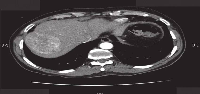
CT Scan of the Abdomen Triphasic Liver A CT scan of the abdomen triphasic liver is a specialized medical imaging test that allows doctors to evaluate the liver and surrounding organs in great detail. This test combines X-ray technology with computer processing to create highly-detailed images of the liver, which can be used to diagnose and monitor a variety of liver diseases, including cancer, cirrhosis, and hepatitis. In this blog post, we will discuss how this test is conducted, what are the common symptoms for ordering the test, how to prepare for the test, how long does it take, and how the results are interpreted. Test procedure and preparation : Before the CT scan, the patient may be asked to remove any metal objects, such as jewelry or dentures, and to wear a hospital gown. The patient will then lie down on a table that slides into a large, doughnut-shaped machine that emits X-rays. The machine will take a series of cross-sectional images of the abdomen, which will be processed by a computer to create detailed, 3D images of the liver. During the scan, the patient may be asked to hold their breath or to stay still to avoid any blurring of the images. In some cases, the doctor may require the use of contrast dye, which helps highlight the blood vessels in and around the liver. The contrast dye may be given intravenously, and the patient may be asked to fast for several hours before the test to avoid any interference with the absorption of the dye. Common symptoms for ordering the test : A CT scan of the abdomen triphasic liver may be ordered by a doctor if the patient is experiencing symptoms such as abdominal pain, bloating, nausea, vomiting, fever, fatigue, or unexplained weight loss. This test may also be used to monitor the progression of a known liver condition or to evaluate the effectiveness of a treatment. Time taken for the test and results interpretation : The duration of the CT scan of the abdomen triphasic liver depends on the complexity of the images required and whether or not a contrast dye is used. Typically, the test takes between 30 minutes to an hour to complete. After the test, the images will be reviewed by a radiologist, who will provide a report to the doctor. The doctor will then discuss the results with the patient, which may include further testing, treatment, or referral to a specialist. A CT scan of the abdomen triphasic liver may be included in a comprehensive health checkup, which is a series of medical tests and screenings that are performed to evaluate a person’s overall health and well-being. A health checkup may also include blood tests, urine tests, electrocardiograms, and other imaging tests, such as a mammogram or a bone density scan. In the context of corporate health and wellness, a CT scan of the abdomen triphasic liver may be offered as part of an employee health program, which aims to promote the health and productivity of the workforce. This program may include regular health screenings, wellness coaching, fitness classes, and other health-related services. In conclusion, a CT scan of the abdomen triphasic liver is a valuable diagnostic tool that can help doctors diagnose and monitor a variety of liver diseases. If you are experiencing any symptoms that may indicate a problem in the liver, or if you are due for a routine health checkup, speak with your doctor about whether a CT scan may be appropriate for you. “Understanding Triphasic Liver CT Scans: Procedure, Benefits, and Applications Introduction: Triphasic liver CT scans are advanced imaging studies used to evaluate liver anatomy and function, providing detailed information about blood supply, tumor characteristics, and overall liver health. In this comprehensive guide, we’ll explore the procedure, benefits, and diverse applications of triphasic liver CT scans, shedding light on their importance in diagnosing and managing various liver conditions. Procedure of Triphasic Liver CT Scan: A triphasic liver CT scan involves three distinct phases: arterial, portal venous, and delayed phases. During the procedure, contrast material is injected intravenously, followed by sequential imaging at specific time intervals to capture the dynamic enhancement patterns of liver tissues and blood vessels. Radiologists carefully analyze the images obtained in each phase to assess liver morphology, detect abnormalities, and evaluate blood flow dynamics. Benefits of Triphasic Liver CT Scan: Triphasic liver CT scans offer several key benefits for diagnosing and managing liver conditions: High Sensitivity for Tumor Detection: By capturing multiple phases of contrast enhancement, triphasic CT scans provide superior sensitivity for detecting liver tumors, including hepatocellular carcinoma (HCC), liver metastases, and other focal lesions. Comprehensive Evaluation of Liver Vasculature: The arterial, portal venous, and delayed phases of the scan allow for detailed assessment of liver vasculature, including arterial blood supply, portal venous flow, and hepatic venous drainage. Accurate Staging and Characterization of Liver Lesions: Triphasic CT scans facilitate accurate staging and characterization of liver lesions, helping clinicians differentiate between benign and malignant tumors, assess tumor vascularity, and plan appropriate treatment strategies. Evaluation of Liver Function and Parenchymal Abnormalities: By visualizing liver enhancement patterns and parenchymal abnormalities in different phases, triphasic CT scans provide valuable insights into liver function, perfusion, and tissue composition. Guided Biopsy and Interventional Procedures: Triphasic CT scans can guide liver biopsy and interventional procedures by precisely localizing target lesions, identifying optimal biopsy sites, and minimizing the risk of complications. Applications of Triphasic Liver CT Scan: Triphasic liver CT scans are utilized for various diagnostic and therapeutic purposes, including: Screening and surveillance for hepatocellular carcinoma (HCC) in high-risk individuals, such as patients with cirrhosis or chronic hepatitis B/C. Detection and characterization of liver metastases from primary malignancies in other organs. Evaluation of portal vein thrombosis, hepatic artery variants, and biliary abnormalities. Assessment of liver abscesses, cysts, hemangiomas, and other focal lesions. Monitoring treatment response and disease progression in patients undergoing liver-directed therapies, such as transarterial chemoembolization (TACE) or radiofrequency ablation (RFA). Interpretation and Follow-Up Recommendations: Interpretation of
Read More
CT Scan of the Abdomen and Pelvis A CT scan of the abdomen and pelvis is a medical imaging test that combines X-ray technology with computer processing to create detailed images of the internal organs and tissues in these regions. This diagnostic test can help doctors detect a wide range of conditions, such as tumors, infections, inflammation, or injuries, that may affect the organs in the abdomen and pelvis, including the liver, kidneys, pancreas, spleen, bladder, and reproductive organs. In this blog post, we will discuss how this test is conducted, what are the common symptoms for ordering the test, how to prepare for the test, how long does it take, and how the results are interpreted. Test procedure and preparation : Before the CT scan, the patient may be asked to remove any metal objects, such as jewelry or dentures, and to wear a hospital gown. The patient will then lie down on a table that slides into a large, doughnut-shaped machine that emits X-rays. The machine will take a series of cross-sectional images of the abdomen and pelvis, which will be processed by a computer to create detailed, 3D images. During the scan, the patient may be asked to hold their breath or to stay still to avoid any blurring of the images. In some cases, the doctor may require the use of contrast dye, which helps highlight the organs and blood vessels in the abdomen and pelvis. The contrast dye may be given orally, intravenously, or through an enema. Before the test, the patient may be asked to fast for several hours, to avoid eating or drinking anything that may interfere with the absorption of the dye. Common symptoms for ordering the test : A CT scan of the abdomen and pelvis may be ordered by a doctor if the patient is experiencing symptoms such as abdominal pain, bloating, diarrhea, constipation, vomiting, blood in the urine or stool, or unexplained weight loss. This test may also be used to evaluate the effectiveness of a treatment for a known condition, to monitor the progression of a disease, or to screen for certain cancers, such as colon or ovarian cancer. Time taken for the test and results interpretation : The duration of the CT scan of the abdomen and pelvis depends on the complexity of the images required and whether or not a contrast dye is used. Typically, the test takes between 30 minutes to an hour to complete. After the test, the images will be reviewed by a radiologist, who will provide a report to the doctor. The doctor will then discuss the results with the patient, which may include further testing, treatment, or referral to a specialist. A CT scan of the abdomen and pelvis may be included in a comprehensive health checkup, which is a series of medical tests and screenings that are performed to evaluate a person’s overall health and well-being. A health checkup may also include blood tests, urine tests, electrocardiograms, and other imaging tests, such as a mammogram or a bone density scan. In the context of corporate health and wellness, a CT scan of the abdomen and pelvis may be offered as part of an employee health program, which aims to promote the health and productivity of the workforce. This program may include regular health screenings, wellness coaching, fitness classes, and other health-related services. In conclusion, a CT scan of the abdomen and pelvis is a valuable diagnostic tool that can help doctors detect and diagnose a wide range of conditions affecting the organs in these regions. If you are experiencing any symptoms that may indicate a problem in the abdomen or pelvis, or if you are due for a routine health checkup, speak with your doctor about whether a CT scan may be appropriate for you. “Comprehensive Guide to CT Scan of Abdomen and Pelvis: Procedure, Benefits, and Applications Introduction: A CT scan of the abdomen and pelvis is a valuable diagnostic tool used to evaluate various abdominal and pelvic conditions. This comprehensive guide provides insights into the importance, procedure, benefits, and applications of CT scans in assessing abdominal and pelvic health. Importance of CT Scan of Abdomen and Pelvis: CT scans of the abdomen and pelvis play a crucial role in diagnosing and evaluating a wide range of medical conditions affecting these regions. From detecting kidney stones and appendicitis to staging tumors and assessing traumatic injuries, CT scans provide detailed images that aid in accurate diagnosis and treatment planning. Procedure for CT Scan of Abdomen and Pelvis: Before the CT scan, patients may be required to fast for a few hours and refrain from drinking fluids to ensure optimal imaging quality. During the procedure, the patient lies on a table that moves through a doughnut-shaped scanner. Contrast material may be injected intravenously to enhance visualization of certain structures. Patients are advised to remain still during the scan to obtain clear images. Precautions and Preparation: Patients should inform their healthcare provider about any allergies, pre-existing medical conditions, or medications they are taking, especially if they have kidney problems or diabetes. It’s essential to follow all instructions provided by the healthcare team regarding fasting, fluid intake, and medication adjustments before the scan. Results Interpretation: After the CT scan, radiologists interpret the images to identify any abnormalities or signs of disease. The results are typically shared with the referring physician, who will discuss the findings with the patient and recommend further diagnostic tests or treatment options if necessary. CT Scan of Abdomen and Pelvis vs. MRI: While both CT scans and MRI provide detailed images of the abdomen and pelvis, they use different imaging techniques. CT scans use X-rays to create cross-sectional images, making them ideal for detecting bone abnormalities and evaluating solid organs. On the other hand, MRI uses magnetic fields and radio waves to produce images, offering superior soft tissue contrast and avoiding ionizing radiation. Benefits of CT Scan of Abdomen and Pelvis: Rapid and non-invasive imaging High-resolution images for accurate
Read More
CT scan of the abdomen A CT scan of the abdomen is a medical imaging test that uses X-rays and computer technology to create detailed images of the abdominal area, including organs such as the liver, pancreas, spleen, and kidneys. It is a non-invasive procedure that can help diagnose and monitor a variety of medical conditions. The test is conducted using a specialized machine called a CT scanner. During the procedure, the patient lies on a table that slides into a donut-shaped machine. The scanner rotates around the patient, taking multiple X-ray images from different angles. These images are then processed by a computer to create detailed images of the abdomen. In terms of preparation for the test, there may be specific instructions given by the healthcare provider or imaging facility. For example, patients may be asked to avoid eating or drinking for a certain period of time before the test, or to avoid certain medications. It’s important to follow any instructions provided to ensure the most accurate results. There are a variety of symptoms and conditions that may warrant a CT scan of the abdomen. For example, the test may be ordered to diagnose or monitor a variety of conditions, including abdominal pain, unexplained weight loss, digestive issues, or blood in the stool. The test may also be used to monitor the progression of certain conditions, such as cancer. The time it takes to complete a CT scan of the abdomen can vary depending on the complexity of the imaging needed. In general, the test itself takes only a few minutes, although additional time may be needed for preparation and positioning. Interpreting the results of a CT scan of the abdomen requires specialized training and expertise. The images produced by the test are highly detailed, and it takes a trained healthcare professional to understand what the images show and how they relate to a patient’s specific medical condition. In many cases, the healthcare provider who ordered the test will review the results with the patient and provide an interpretation. In the context of health checkups and wellness programs, CT scans of the abdomen can be a valuable tool for monitoring overall health and identifying potential health issues before they become more serious. Many wellness programs offer CT scans of the abdomen as part of a comprehensive health evaluation, which can help individuals take a proactive approach to their health. Corporate health programs may also incorporate CT scans of the abdomen as a way to monitor the health of employees and identify potential health risks. For example, employers may offer CT scans of the abdomen as part of a preventative health screening program, or as part of a wellness incentive program. By providing employees with access to these types of imaging tests, employers can help employees take a more active role in their health and potentially reduce healthcare costs over the long term. In conclusion, a CT scan of the abdomen is a non-invasive medical imaging test that can help diagnose and monitor a variety of medical conditions. The test is conducted using a specialized machine called a CT scanner, and may require specific preparation depending on the body part being examined. Symptoms that may prompt a healthcare provider to order a CT scan of the abdomen include abdominal pain, unexplained weight loss, digestive issues, or blood in the stool. Interpreting the results of a CT scan of the abdomen requires specialized training and expertise, and the test can be a valuable tool for monitoring overall health and identifying potential health issues before they become more serious.
Read More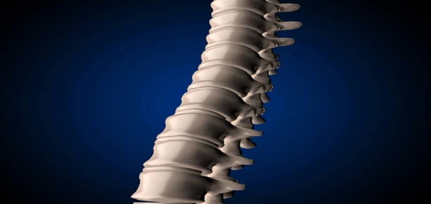
CT Scan Spiral Cervical Spine A CT (computed tomography) scan spiral cervical spine is a medical imaging test used to examine the cervical spine or neck area. It is a non-invasive procedure that uses X-rays and computer technology to create detailed images of the cervical spine. The test is conducted by having the patient lie on a table that moves through a large, circular machine. During the scan, the machine rotates around the patient, taking multiple X-ray images from different angles. A computer then combines these images to create a detailed 3D image of the cervical spine. There is usually no specific preparation required for a CT scan spiral cervical spine. However, patients may be asked to remove any metal objects such as jewelry, eyeglasses, and dentures before the scan, as they can interfere with the imaging. Patients may also be asked to wear a hospital gown and to avoid eating or drinking for a few hours prior to the test. A CT scan spiral cervical spine may be ordered by a doctor if a patient is experiencing symptoms such as neck pain, weakness, numbness or tingling in the arms, or difficulty walking. The test may also be ordered as part of a routine health checkup or wellness program, particularly for individuals with a history of spinal cord injury or degenerative conditions such as arthritis. Additionally, a CT scan spiral cervical spine may be used as part of a corporate health program to assess the health of employees and identify potential health risks. The time taken for a CT scan spiral cervical spine can vary depending on the individual case and the imaging equipment used. Generally, the test takes between 30 and 60 minutes to complete. Patients may be asked to hold their breath for a few seconds during the scan to prevent blurring of the images. The results of a CT scan spiral cervical spine are typically interpreted by a radiologist, who will analyze the images and look for any abnormalities or signs of disease. The images can reveal a range of conditions, including herniated discs, spinal stenosis, fractures, and tumors. If a significant abnormality is detected, further testing or treatment may be required. In conclusion, a CT scan spiral cervical spine is a valuable diagnostic tool for examining the neck area and its structures. It is a non-invasive procedure that can be used to diagnose a range of conditions and is often ordered as part of routine health checkups, wellness programs, and corporate health assessments. While there is usually no specific preparation required for the test, patients should follow any instructions provided by their doctor or imaging facility to ensure the best possible imaging results.
Read More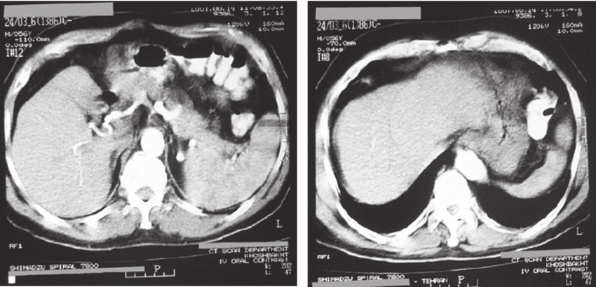
CT Scan Spiral Pelvis A CT Scan Spiral Pelvis is a diagnostic medical test that uses X-rays to create detailed images of the pelvis area, including the hip bones, pelvic organs, blood vessels, and surrounding soft tissues. This test can help diagnose a wide range of conditions affecting the pelvis, such as fractures, tumors, infections, and other abnormalities. In this article, we will discuss the various aspects of a CT Scan Spiral Pelvis, including the test procedure, preparation, common symptoms, time taken for the test, result interpretation, and its importance in corporate health wellness packages. Test Procedure : During a CT Scan Spiral Pelvis, the patient lies down on a table that slides into a cylindrical machine. The machine uses X-rays to take multiple images of the pelvis area from different angles. These images are processed by a computer to create a detailed 3D image of the pelvis area. The entire process takes around 30-45 minutes. Test Preparation : Before a CT Scan Spiral Pelvis, patients may be asked to avoid eating or drinking for several hours before the test. Patients may also be asked to remove any metal objects, jewelry, or clothing that might interfere with the X-rays. In some cases, patients may need to drink a contrast solution before the test to help highlight certain structures in the pelvis. Common Symptoms for Ordering the Test : A CT Scan Spiral Pelvis is usually ordered by doctors when a patient has symptoms such as pelvic pain, hip pain, difficulty urinating, blood in the urine, or other signs of a possible pelvic condition. The test may also be ordered to evaluate the extent of an injury or to monitor the progression of a known condition, such as a tumor. Time Taken for the Test and its Results Interpretation : A CT Scan Spiral Pelvis usually takes around 30-45 minutes. The images generated by the test are interpreted by a radiologist, who will then provide a report to the referring physician. The report will contain information about any abnormalities found in the pelvis area, as well as recommendations for further treatment or testing if necessary. Importance in Corporate Health Wellness Packages : A CT Scan Spiral Pelvis can be an important component of a corporate health wellness package. By offering this test to employees, companies can help identify potential health issues early, which can lead to more effective treatment and better outcomes. Additionally, offering health screenings and tests can help improve employee morale and productivity by demonstrating that the company is invested in the well-being of its employees. In conclusion, a CT Scan Spiral Pelvis is a valuable diagnostic tool that can help diagnose and evaluate a range of conditions affecting the pelvis area. The test is generally non-invasive, although some patients may need to drink a contrast solution before the test. If you are experiencing symptoms such as pelvic pain, hip pain, or difficulty urinating, talk to your doctor about whether a CT Scan Spiral Pelvis may be appropriate for you. Additionally, if you are a business owner or HR professional, consider offering health screenings and tests, such as a CT Scan Spiral Pelvis, as part of your corporate wellness program to promote the health and well-being of your employees.
Read More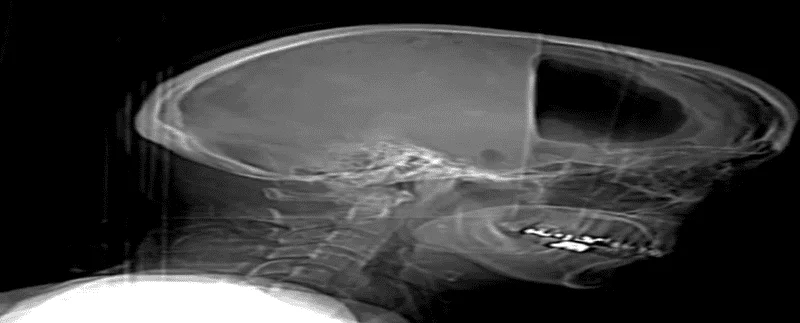
CT Scan Spiral Face A CT scan spiral face, also known as a facial CT scan, is a diagnostic imaging test that uses X-rays and computer technology to produce detailed images of the bones, muscles, and other tissues of the face. It is a non-invasive procedure that provides cross-sectional images of the face, allowing doctors to diagnose a wide range of conditions. The test is typically conducted in a hospital or diagnostic imaging center. Before the procedure, patients are asked to remove any metal objects, such as jewelry, watches, and hairpins, as these can interfere with the X-rays. Patients may also be asked to change into a hospital gown or other loose-fitting clothing. During the test, the patient lies down on a narrow table that slides into a large, doughnut-shaped machine. The machine rotates around the patient, taking multiple X-ray images from different angles. These images are then processed by a computer to create detailed, three-dimensional images of the face. Preparation for a facial CT scan: Patients may be asked to fast for a period of time before the procedure. This is to ensure that the stomach and intestines are empty, which can improve the clarity of the images. Patients may also be asked to avoid taking certain medications or supplements before the procedure, as these can affect the results. Common symptoms: The most common symptoms that may lead to a facial CT scan include facial pain, swelling, or deformity, as well as chronic sinus infections, or a history of facial trauma. The length of time that a facial CT scan takes: Depending on a number of factors, such as the size of the area being imaged and the quality of the images needed. Typically, the test can take between 10 and 30 minutes to complete. After the test is complete, a radiologist will review the images and provide a report to the patient’s doctor. The report will include information about any abnormalities that were found, as well as recommendations for further testing or treatment if necessary. A facial CT scan can be an important part of a health checkup, wellness program, or corporate health initiative. By identifying potential health issues early on, patients can take steps to address them and improve their overall health and wellbeing. Additionally, regular facial CT scans may be recommended for patients with a history of facial trauma or chronic sinus infections. Conclusion: A facial CT scan is a non-invasive diagnostic test that uses X-rays and computer technology to produce detailed images of the bones, muscles, and other tissues of the face. The test can be conducted in a hospital or imaging center, and patients may be asked to fast or avoid certain medications before the procedure. Common symptoms that may lead to a facial CT scan include facial pain, swelling, or deformity, as well as chronic sinus infections or a history of facial trauma. The length of the test can vary, and results are typically provided to the patient’s doctor for further evaluation and treatment. By incorporating regular facial CT scans into health checkups, wellness programs, and corporate health initiatives, patients can take proactive steps to maintain their health and wellbeing.
Read More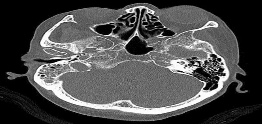
CT Scan of the Temporal Bone or Mastoid Bone A CT Scan of the Temporal Bone or Mastoid Bone is a diagnostic medical imaging test used to evaluate the ear, including the ear canal, middle ear, and mastoid bone. This imaging test provides detailed images of the bone structure and surrounding soft tissues of the ear. In this article, we will discuss the procedure of the CT Scan of Temporal or Mastoid Bone, test preparation, common symptoms, time taken for the test, result interpretation, and its importance in corporate health wellness packages. Test Procedure: During a CT Scan of Temporal or Mastoid Bone, the patient is positioned on a table that slides through a doughnut-shaped machine. The machine rotates around the patient, taking multiple images of the ear and the surrounding structures. The images captured are then processed by a computer to produce detailed images of the ear and its components. This test usually takes about 15-30 minutes. Test Preparation: Before the CT Scan of Temporal or Mastoid Bone, the patient should wear comfortable, loose-fitting clothing without metal fasteners or jewelry that may interfere with the imaging. Patients should also inform their physician of any pre-existing allergies or medical conditions that may affect the CT scan. They may also be asked to remove any removable dental work, such as dentures, as it may obstruct the imaging. Common Symptoms for Ordering the Test: A CT Scan of Temporal or Mastoid Bone may be ordered by a doctor to investigate the following symptoms or conditions: Chronic ear infections Hearing loss Tinnitus (ringing in the ears) Vertigo (dizziness) Facial weakness or paralysis Mastoiditis (infection of the mastoid bone) Cholesteatoma (an abnormal skin growth behind the eardrum) Trauma or injury to the ear or head Time Taken for the Test and its Results Interpretation The CT Scan of Temporal or Mastoid Bone usually takes about 15-30 minutes. The results of the test are evaluated by a radiologist, who will interpret the images and provide a report to the referring physician. The report will include information about the condition of the ear, including any abnormal structures, infections, or growths. Importance in Corporate Health Wellness Packages: A CT Scan of Temporal or Mastoid Bone is a crucial diagnostic tool for individuals who are experiencing symptoms related to their ear or surrounding structures. By offering this test as part of corporate health wellness packages, companies can provide their employees with the opportunity to detect ear-related conditions early, and promote early treatment to prevent potential complications. Additionally, regular health screenings and tests can help in identifying potential health concerns early and provide a preventative approach to healthcare. Conclusion: CT Scan of Temporal or Mastoid Bone is a safe, non-invasive diagnostic imaging test used to evaluate the ear and surrounding structures. The test provides detailed images of the bone and surrounding soft tissue structures. Patients do not need to prepare significantly for this test, but they should inform their doctor about any medical conditions or allergies they have before the test. The results of the test are interpreted by a radiologist, and the report is sent to the referring physician. Companies can offer CT Scan of Temporal or Mastoid Bone as part of their corporate wellness packages to promote the well-being of their employees by enabling early detection and timely treatment of ear-related conditions.
Read More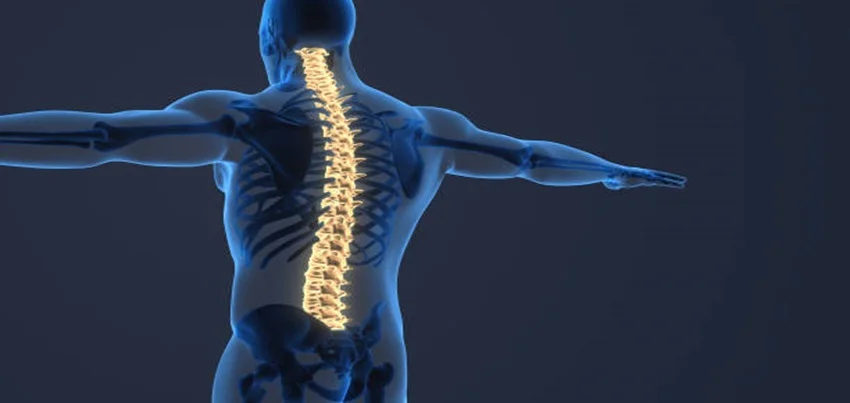
CT Scan Spiral Whole Spine A CT Scan Spiral Whole Spine is a medical test that uses X-rays to create detailed images of the entire spine, from the neck to the lower back. This test can help diagnose a wide range of conditions affecting the spine, such as herniated discs, spinal stenosis, fractures, tumors, and other abnormalities. In this article, we will discuss the various aspects of a CT Scan Spiral Whole Spine, including the test procedure, preparation, common symptoms, time taken for the test, result interpretation, and its importance in corporate health wellness packages. Test Procedure: During a CT Scan Spiral Whole Spine, the patient lies down on a table that slides into a cylindrical machine. The machine uses X-rays to take multiple images of the spine from different angles. These images are processed by a computer to create a detailed 3D image of the entire spine. The entire process takes around 30-45 minutes. Test Preparation: Before a CT Scan Spiral Whole Spine, patients may be asked to avoid eating or drinking for several hours before the test. Patients may also be asked to remove any metal objects, jewelry, or clothing that might interfere with the X-rays. In some cases, patients may need to drink a contrast solution before the test to help highlight certain structures in the spine. Common Symptoms for Ordering the Test: A CT Scan Spiral Whole Spine is usually ordered by doctors when a patient has symptoms such as back pain, numbness or tingling in the arms or legs, weakness in the limbs, or other signs of a possible spine condition. The test may also be ordered to evaluate the extent of an injury or to monitor the progression of a known condition, such as a tumor. Time Taken for the Test and its Results Interpretation: A CT Scan Spiral Whole Spine usually takes around 30-45 minutes. The images generated by the test are interpreted by a radiologist, who will then provide a report to the referring physician. The report will contain information about any abnormalities found in the spine, as well as recommendations for further treatment or testing if necessary. Importance in Corporate Health Wellness Packages: A CT Scan Spiral Whole Spine can be an important component of a corporate health wellness package. By offering this test to employees, companies can help identify potential health issues early, which can lead to more effective treatment and better outcomes. Additionally, offering health screenings and tests can help improve employee morale and productivity by demonstrating that the company is invested in the well-being of its employees. In conclusion, a CT Scan Spiral Whole Spine is a valuable diagnostic tool that can help diagnose and evaluate a range of conditions affecting the entire spine. The test is generally non-invasive, although some patients may need to drink a contrast solution before the test. If you are experiencing symptoms such as back pain, numbness or tingling in the arms or legs, or weakness in the limbs, talk to your doctor about whether a CT Scan Spiral Whole Spine may be appropriate for you. Additionally, if you are a business owner or HR professional, consider offering health screenings and tests, such as a CT Scan Spiral Whole Spine, as part of your corporate wellness program to promote the health and well-being of your employees.
Read MoreTop rated products
-
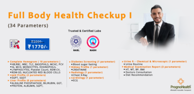 Full Body Health Checkup I
Original price was: ₹3,000.00.₹1,770.00Current price is: ₹1,770.00.
Full Body Health Checkup I
Original price was: ₹3,000.00.₹1,770.00Current price is: ₹1,770.00.
-
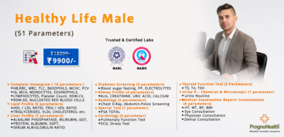 Healthy Life Male
Original price was: ₹18,000.00.₹9,900.00Current price is: ₹9,900.00.
Healthy Life Male
Original price was: ₹18,000.00.₹9,900.00Current price is: ₹9,900.00.
-
 Complete Men’s Health Profile – 40+ – 136 Parameters
₹2,899.00
Complete Men’s Health Profile – 40+ – 136 Parameters
₹2,899.00
-
 OFFICE STAFF PACK II
Original price was: ₹2,400.00.₹1,320.00Current price is: ₹1,320.00.
OFFICE STAFF PACK II
Original price was: ₹2,400.00.₹1,320.00Current price is: ₹1,320.00.
-
 Full Body Health Checkup III
₹11,000.00
Full Body Health Checkup III
₹11,000.00