PrognoHealth – Corporate Health & Wellness Specialist


CT scan of a joint A CT scan of a joint is a test that provides images of a specific joint in the body. This test is commonly ordered when a patient is experiencing symptoms related to a joint. Test Conducted and Preparation:During a CT scan of a joint, the patient lies down on a table that slides into the CT machine. The machine takes multiple images from different angles and uses computer processing to create a 3D image of the joint. The test is painless and usually takes less than 30 minutes to complete. There is no specific preparation required for a CT scan of a joint. However, patients may be asked to remove any metal objects, such as jewelry, before the test. Common Symptoms for Ordering the Test: A CT scan of a joint may be ordered by a doctor if a patient is experiencing symptoms such as: Pain or stiffness in the joint Limited range of motion in the joint Swelling or inflammation in the joint Trauma to the joint Suspected fractures or dislocations Time Taken for the Test and its Results Interpretations: The test itself takes only a few minutes, but the entire process, including check-in, preparation, and post-test consultation, may take up to an hour. After the test, a radiologist will review the images and prepare a report for the patient’s doctor. The doctor will then discuss the results with the patient and provide any necessary treatment recommendations. A CT scan of a joint may be included as part of a comprehensive health checkup or wellness program. It can help detect and diagnose conditions such as fractures, dislocations, and arthritis. For corporate health programs, this test may be used to assess employees’ health risks and provide preventive care. Conclusion: CT scan of a joint is a valuable diagnostic tool that can provide detailed images of a specific joint in the body. It is a quick and painless test that can help diagnose a range of conditions related to the joint. This test may be included as part of a health checkup or wellness program and can be useful in corporate health settings to assess and manage employee health risks.
Read More
Sodium Test A sodium test is a laboratory test that measures the level of sodium in the blood. Sodium is an electrolyte that plays a crucial role in maintaining the balance of fluids in the body and in regulating the function of muscles and nerves. Pre-test preparation: No special preparation is typically required for a sodium test. Testing method: A small sample of blood is taken from a vein in the arm and sent to a laboratory for analysis. The sample is usually analyzed using a device called an electrolyte analyzer. Common symptoms for prescribing this test: A sodium test may be ordered if a person has symptoms of a condition that affects the level of sodium in the blood, such as dehydration, kidney disease, or certain types of heart and lung disorders. It can also be used as a part of routine lab work to check overall health status. Diagnosis: The results of a sodium test are used to help diagnose and monitor a variety of conditions, including dehydration, kidney disease, and certain types of heart and lung disorders. Elevated levels of sodium can indicate dehydration, while low levels can indicate kidney disease, certain types of heart and lung disorders, or the use of certain medications. Reference range: The reference range for sodium levels varies depending on the lab and the population being tested, but generally, a normal range is 135 to 145 mEq/L. Normal values: Sodium levels are normally between 135 to 145 mEq/L. Medical disclaimer: The above information is provided for general educational purposes and is not intended to be a substitute for professional medical advice, diagnosis, or treatment. Always seek the advice of your physician or other qualified healthcare provider with any questions you may have regarding a medical condition. It is important to note that test results should be always interpreted in the context of a patient’s clinical presentation and other test results, and treatment should be determined by a healthcare professional.
Read More
Zinc Test A zinc test is a laboratory test that measures the level of zinc in the blood. Zinc is an essential mineral that plays a vital role in many bodily functions, including growth and development, immune function, and wound healing. Pre-test preparation: No special preparation is typically required for a zinc test. Testing method: A small sample of blood is taken from a vein in the arm and sent to a laboratory for analysis. The sample is usually analyzed using a device called an atomic absorption spectrophotometer. Common symptoms for prescribing this test: A zinc test may be ordered if a person has symptoms of a condition that affects the level of zinc in the blood, such as malnutrition, malabsorption, or certain types of liver or kidney disease. It can also be used to monitor treatment of zinc deficiency or to detect early onset of zinc deficiency. Diagnosis: The results of a zinc test are used to help diagnose and monitor a variety of conditions, including zinc deficiency, malnutrition, malabsorption, and certain types of liver or kidney disease. Elevated levels of zinc can indicate liver disease, while low levels can indicate zinc deficiency, malnutrition, or malabsorption. Reference range: The reference range for zinc levels varies depending on the lab and the population being tested, but generally, a normal range is 70 to 150 mcg/dL. Normal values: Zinc levels are normally between 70 to 150 mcg/dL. Medical disclaimer: The above information is provided for general educational purposes and is not intended to be a substitute for professional medical advice, diagnosis, or treatment. Always seek the advice of your physician or other qualified healthcare provider with any questions you may have regarding a medical condition. It is important to note that test results should be always interpreted in the context of a patient’s clinical presentation and other test results, and treatment should be determined by a healthcare professional.
Read More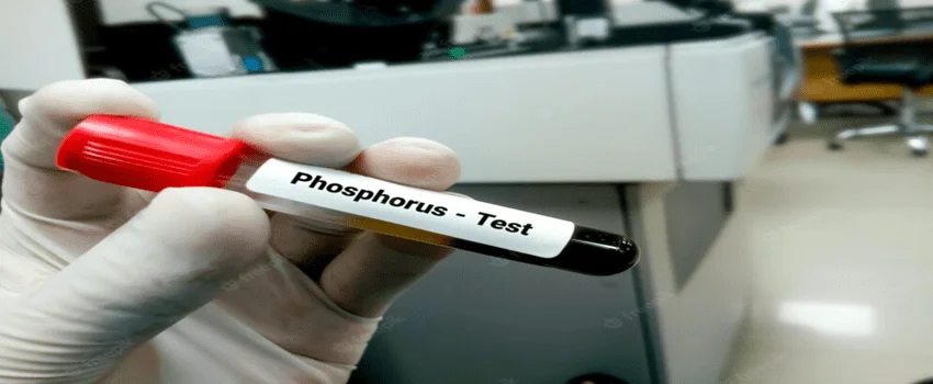
Phosphorus is an essential mineral that plays an important role in many bodily functions such as maintaining strong bones and teeth, filtering waste products from the kidneys, and helping cells and organs to function properly.
Read More
CT scan of the orbit A CT (computed tomography) scan of the orbit is a diagnostic imaging test that uses X-rays and computer technology to produce detailed images of the eye socket area. This test is commonly ordered when a patient is experiencing symptoms related to the eyes or surrounding area. Test Conducted and Preparation: During a CT scan of the orbit, the patient lies down on a table that slides into the CT machine. The machine takes multiple images from different angles and uses computer processing to create a 3D image of the eye socket area. The test is painless and usually takes less than 30 minutes to complete. There is no specific preparation required for a CT scan of the orbit. However, patients may be asked to remove any metal objects, such as jewelry or eyeglasses, before the test. Common Symptoms for Ordering the Test: A CT scan of the orbit may be ordered by a doctor if a patient is experiencing symptoms such as:Pain or discomfort in the eyes or surrounding areaSwelling or lumps around the eyesDouble vision or blurred visionDecreased vision or loss of visionEye discharge or crustingEye injuries or traumaThese symptoms could indicate conditions such as infections, tumors, or injuries to the eye socket area. Time Taken for the Test and its Results Interpretations: The test itself takes only a few minutes, but the entire process, including check-in, preparation, and post-test consultation, may take up to an hour. After the test, a radiologist will review the images and prepare a report for the patient’s doctor. The doctor will then discuss the results with the patient and provide any necessary treatment recommendations. A CT scan of the orbit may be included as part of a comprehensive health checkup or wellness program. It can help detect and diagnose conditions such as infections, tumors, and injuries to the eye socket area. For corporate health programs, this test may be used to assess employees’ health risks and provide preventive care. In addition, a CT scan of the orbit can be useful for monitoring the effectiveness of treatments for conditions such as tumors or infections. It can also be used to guide surgical procedures or biopsies of the eye socket area. While a CT scan of the orbit is generally considered a safe and effective diagnostic tool, it does involve exposure to ionizing radiation. Therefore, doctors typically limit the number of CT scans a patient receives to reduce the risk of radiation-related side effects. Patients should always discuss the risks and benefits of any imaging test with their doctor. In conclusion, a CT scan of the orbit is a valuable diagnostic tool that can provide detailed images of the eye socket area. It is a quick and painless test that can help diagnose a range of conditions related to the eyes or surrounding area. This test may be included as part of a health checkup or wellness program and can be useful in corporate health settings to assess and manage employee health risks. It is important for patients to discuss the risks and benefits of any imaging test with their doctor and to follow any preparation instructions provided prior to the test.
Read More
Magnetic Resonance Cholangiopancreatography (MRCP) is a diagnostic imaging test that uses magnetic fields and radio waves to produce detailed images of the bile ducts and pancreas
Read More
MRI of the orbit Magnetic Resonance Imaging (MRI) of the orbit is a diagnostic test that uses a powerful magnetic field, radio waves, and a computer to produce detailed images of the eyes and surrounding structures. The test is conducted to evaluate a range of conditions affecting the eyes and vision. Test Conducted: During an MRI of the orbit, the patient lies on a table that slides into a cylindrical machine that houses the MRI scanner. The scanner uses a magnetic field and radio waves to create images of the eyes and surrounding structures. The test typically takes between 30 and 60 minutes to complete, depending on the complexity of the exam and the patient’s ability to remain still during the procedure. Test Preparation: For an MRI of the orbit, patients should wear comfortable, loose-fitting clothing without metal zippers or buttons. The patient will be asked to remove any metal objects, such as jewelry, watches, or belts. The patient may also be asked to fast for a few hours before the exam, depending on the reason for the test. Common Symptoms for Ordering the Test: MRI of the orbit may be ordered by a physician if a patient has symptoms such as vision loss or changes, double vision, pain in the eyes, or abnormal bulging of the eyes. The test can help diagnose a range of conditions affecting the eyes and surrounding structures, including tumors, inflammation, infections, and other abnormalities. Time Taken for the Test and Interpretation of Results: An MRI of the orbit typically takes between 30 and 60 minutes to complete, and patients can return to their normal activities immediately after the test. The images produced by the MRI are examined by a radiologist, who will interpret the results and provide a report to the ordering physician. The physician will then review the results with the patient and determine the appropriate course of treatment based on the findings. Using Health Checkup, Wellness and Corporate Health as Key Words: MRI of the orbit is an important tool for diagnosing and treating a range of conditions affecting the eyes and vision, and can be used in regular health checkups and corporate wellness programs. Early detection of eye problems is critical to maintaining good vision and overall health. In addition, some corporate health programs offer wellness screenings that include eye exams, and an MRI of the orbit may be ordered as part of a comprehensive wellness evaluation. In conclusion, MRI of the orbit is a valuable diagnostic tool that can help detect a range of eye conditions that can affect overall health and wellness. The test is non-invasive and typically takes between 30 and 60 minutes to complete, with some preparation required such as removing metal objects and fasting for a few hours before the exam. The results are interpreted by a radiologist and reviewed by the ordering physician, who will determine the appropriate course of treatment based on the findings. Regular health checkups and corporate wellness programs can also incorporate eye exams, including MRI of the orbit, to help detect and manage eye problems.
Read More
Pregnancy-associated plasma protein A (PAPP-A) is a protein that is produced by the placenta during pregnancy. The PAPP-A test is a blood test that measures the level of PAPP-A in a pregnant woman’s blood.
Read More
Liver Function Test (LFT) A Liver Function Test (LFT) is a group of blood tests that are used to evaluate the functioning of the liver. The liver is responsible for a wide range of functions including detoxification, protein synthesis, and bile production. Pre-test preparation: There is no specific preparation required for a LFT. Testing method: A small sample of blood is taken from a vein in your arm and sent to a laboratory for analysis. The test typically includes measurement of enzymes such as Alanine transaminase (ALT), Aspartate transaminase (AST), Alkaline phosphatase (ALP), Bilirubin, and Albumin. Common symptoms that may prompt your doctor to order a LFT include: jaundice (yellowing of the skin and eyes), abdominal pain, and dark urine. Diagnosis: LFTs are used to detect and monitor liver disease, such as hepatitis, cirrhosis, and liver cancer. Elevated levels of liver enzymes can indicate damage to liver cells, while low levels of albumin can indicate a problem with liver function. Reference range: The normal range for LFTs varies depending on the laboratory that performs the test, but typical ranges for enzymes are:ALT: 0-40 U/LAST: 0-40 U/LALP Liver function tests (LFTs) are a group of blood tests that are used to assess the health of the liver and to diagnose and monitor liver disease or damage.Pre-test preparation typically includes fasting for 8-12 hours before the test, as well as avoiding alcohol for at least 24 hours prior to the test.The test is typically done by drawing blood from a vein in the arm and sending it to a lab for analysis. Common symptoms that may lead a healthcare provider to prescribe LFTs include jaundice (yellowing of the skin and eyes), fatigue, abdominal pain, and loss of appetite.These tests can help diagnose a wide range of liver conditions including hepatitis, cirrhosis, and liver cancer. A common set of LFTs includes:Alanine transaminase (ALT)Aspartate transaminase (AST)Alkaline phosphatase (ALP)Total bilirubinAlbuminProthrombin time (PT)International normalized ratio (INR) Reference ranges and normal values vary depending on the specific test and the lab that is performing the analysis. It’s important to consult with a healthcare provider or the lab that performed the test to understand the results and their implications. It’s important to note that liver function tests are not definitive in diagnosing liver disease, they are just a way to screen the liver and more definitive tests like imaging and biopsy are often required. Additionally, other medical conditions can also affect the results of LFTs, so it is important to consult a healthcare professional for proper interpretation of the results.
Read More
Lactate Dehydrogenase (LDH) Test Lactate Dehydrogenase (LDH) test is a blood test that measures the level of the enzyme lactate dehydrogenase (LDH) in the blood. LDH is found in many body tissues, including the heart, liver, kidneys, muscles, and red blood cells. Pre-test preparation: There is no specific preparation required for a LDH test. Testing method: A small sample of blood is taken from a vein in your arm and sent to a laboratory for analysis. Common symptoms that may prompt your doctor to order a LDH test include: jaundice (yellowing of the skin and eyes), abdominal pain, and dark urine. Diagnosis: Elevated levels of LDH in the blood can indicate damage to organs or tissues such as the heart, liver, or kidneys. It is also used as a marker of certain types of cancer, such as lymphoma, and to monitor treatment for these conditions. Reference range: The normal range for LDH in the blood varies depending on the laboratory that performs the test, but typically ranges from 125-225 U/L. Normal values may vary depending on the laboratory that performs the test. Medical disclaimer: The information provided is not intended to be a substitute for professional medical advice, diagnosis, or treatment. Always seek the advice of your physician or other qualified healthcare provider with any questions you may have regarding a medical condition. It is important to follow the instructions given by your doctor or the laboratory performing the test. If you have symptoms that suggest organ or tissue damage or have been advised to have a LDH test, it is important to work closely with your doctor to understand the results and any further actions that may be necessary to manage your health.
Read MoreTop rated products
-
 Full Body Health Checkup I
Original price was: ₹3,000.00.₹1,770.00Current price is: ₹1,770.00.
Full Body Health Checkup I
Original price was: ₹3,000.00.₹1,770.00Current price is: ₹1,770.00.
-
 Young Life Basic
Original price was: ₹3,000.00.₹1,350.00Current price is: ₹1,350.00.
Young Life Basic
Original price was: ₹3,000.00.₹1,350.00Current price is: ₹1,350.00.
-
 COVID 19 Rapid Antigen Test
Original price was: ₹668.00.₹501.00Current price is: ₹501.00.
COVID 19 Rapid Antigen Test
Original price was: ₹668.00.₹501.00Current price is: ₹501.00.
-
 MFG IND PACK III
Original price was: ₹3,900.00.₹2,145.00Current price is: ₹2,145.00.
MFG IND PACK III
Original price was: ₹3,900.00.₹2,145.00Current price is: ₹2,145.00.
-
 Healthy Life Advance Male
Original price was: ₹15,000.00.₹8,250.00Current price is: ₹8,250.00.
Healthy Life Advance Male
Original price was: ₹15,000.00.₹8,250.00Current price is: ₹8,250.00.