PrognoHealth – Corporate Health & Wellness Specialist

Eye Care in Digital World – Importance of regular eye Checkups Maintaining good vision is essential for navigating daily life, yet many individuals overlook the importance of regular eye checkups until they experience noticeable problems with their vision.

Read More

CT scan spiral Brain + Angio A CT (computed tomography) scan spiral brain + angio is a medical imaging test used to examine the brain and its blood vessels. It is a non-invasive procedure that uses X-rays and computer technology to create detailed images of the brain and its blood vessels. The test is conducted by having the patient lie on a table that moves through a large, circular machine. During the scan, the machine rotates around the patient, taking multiple X-ray images from different angles. A computer then combines these images to create a detailed 3D image of the brain and its blood vessels. There is usually no specific preparation required for a CT scan spiral brain + angio. However, patients may be asked to remove any metal objects such as jewelry, eyeglasses, and dentures before the scan, as they can interfere with the imaging. Patients may also be asked to wear a hospital gown and to avoid eating or drinking for a few hours prior to the test. A CT scan spiral brain + angio may be ordered by a doctor if a patient is experiencing symptoms such as headaches, seizures, dizziness, or changes in vision or hearing. The test may also be ordered as part of a routine health checkup or wellness program, particularly for individuals with a history of stroke or heart disease. Additionally, a CT scan spiral brain + angio may be used as part of a corporate health program to assess the health of employees and identify potential health risks. The time taken for a CT scan spiral brain + angio can vary depending on the individual case and the imaging equipment used. Generally, the test takes between 30 and 60 minutes to complete. Patients may be asked to hold their breath for a few seconds during the scan to prevent blurring of the images. The results of a CT scan spiral brain + angio are typically interpreted by a radiologist, who will analyze the images and look for any abnormalities or signs of disease. The images can reveal a range of conditions, including brain tumors, aneurysms, bleeding in the brain, and narrowing or blockages in blood vessels. If a significant abnormality is detected, further testing or treatment may be required. In conclusion, a CT scan spiral brain + angio is a valuable diagnostic tool for examining the brain and its blood vessels. It is a non-invasive procedure that can be used to diagnose a range of conditions and is often ordered as part of routine health checkups, wellness programs, and corporate health assessments. While there is usually no specific preparation required for the test, patients should follow any instructions provided by their doctor or imaging facility to ensure the best possible imaging results.

Read More

MRI Of The Thoracic Spine Magnetic Resonance Imaging (MRI) of the thoracic spine is a non-invasive diagnostic test that uses a powerful magnet, radio waves, and a computer to produce detailed images of the spinal cord and surrounding tissues. The test is conducted to evaluate the thoracic spine for various conditions that can cause pain, numbness, or weakness in the upper back, chest, abdomen, or legs. Test Conducted:During the MRI of the thoracic spine, the patient lies on a table that slides into a cylindrical machine that houses the MRI scanner. The scanner uses a magnetic field and radio waves to create images of the thoracic spine. The test takes between 30 and 60 minutes to complete, depending on the complexity of the exam and the patient’s ability to remain still during the procedure. Test Preparation:For an MRI of the thoracic spine, patients should wear comfortable, loose-fitting clothing without metal zippers or buttons. The patient will be asked to remove any metal objects, such as jewellery, watches, or belts. The patient will also be asked to complete a screening form to ensure that there are no contraindications for the test. Common Symptoms for Ordering the Test:MRI of the thoracic spine may be ordered by a physician if a patient has symptoms such as pain or weakness in the upper back, chest, or abdomen, difficulty walking, numbness or tingling in the arms, legs, or torso, or signs of a spinal cord injury or tumor. The test can help diagnose a range of conditions affecting the thoracic spine, including herniated discs, spinal cord compression, spinal stenosis, tumors, infections, and other abnormalities. Time Taken for the Test and Interpretation of Results:An MRI of the thoracic spine typically takes between 30 and 60 minutes to complete, and patients can return to their normal activities immediately after the test. The images produced by the MRI are examined by a radiologist, who will interpret the results and provide a report to the ordering physician. The physician will then review the results with the patient and determine the appropriate course of treatment based on the findings. MRI of the thoracic spine is an important tool for diagnosing and treating a range of spinal conditions that can affect overall health and wellness. Regular health checkups can help detect and manage spinal problems before they become more serious. Additionally, many corporate health programs offer wellness screenings that include spinal exams, and an MRI of the thoracic spine may be ordered as part of a comprehensive wellness evaluation. In conclusion, MRI of the thoracic spine is a valuable diagnostic tool that can help detect a range of spinal conditions that can affect overall health and wellness. The test is non-invasive and typically takes between 30 and 60 minutes to complete, with no preparation required other than removing metal objects. The results are interpreted by a radiologist and reviewed by the ordering physician, who will determine the appropriate course of treatment based on the findings. Regular health checkups and corporate wellness programs can also incorporate spinal exams, including MRI of the thoracic spine, to help detect and manage spinal problems. Magnetic Resonance Imaging (MRI) of the thoracic spine is a diagnostic procedure that provides detailed images of the middle segment of the spine, which runs from the base of the neck to the upper part of the lower back. This non-invasive imaging technique is particularly useful for evaluating soft tissues, the spinal cord, intervertebral discs, and surrounding structures Anatomy of the Thoracic Spine The thoracic spine consists of 12 vertebrae, labelled T1 to T12, and forms the middle portion of the spine. Key components include: • Vertebrae: The bony segments that provide structural support and protect the spinal cord.• Intervertebral Discs: Soft cushions between the vertebrae that absorb shock and allow flexibility.• Spinal Cord: The bundle of nerves running through the spinal canal.• Nerve Roots: Branches of nerves exiting the spinal cord.• Facet Joints: Small joints at the back of each vertebra that facilitate movement.• Ligaments and Muscles: Structures that support and stabilise the spine. Why is a Thoracic Spine MRI Done? MRI of the thoracic spine is used to diagnose and monitor various conditions, including: 1. Back Pain:o Persistent or unexplained mid-back pain.o Pain radiating around the chest or abdomen (thoracic radiculopathy).2. Degenerative Diseases:o Herniated Discs: Discs protruding and compressing spinal nerves.o Spinal Stenosis: Narrowing of the spinal canal causing nerve compression.o Osteoarthritis: Degeneration of the spine’s facet joints.3. Spinal Cord Disorders:o Myelopathy: Compression or injury to the spinal cord causing neurological symptoms.o Multiple Sclerosis (MS): Detection of demyelinating lesions in the spinal cord.4. Trauma:o Fractures or dislocations from accidents or injuries.5. Infections and Inflammatory Conditions:o Osteomyelitis: Infection of the bones.o Discitis: Infection of the intervertebral discs.o Ankylosing Spondylitis: Inflammatory disease affecting the spine.6. Tumors:o Primary Tumors: Tumors originating in the spine or spinal cord.o Metastatic Tumors: Cancer that has spread from other parts of the body to the spine.7. Post-Surgical Evaluation:o Monitoring recovery or complications after spinal surgery. How is the Thoracic Spine MRI Performed? 1. Preparation:o Generally, no special preparation is required.o Inform your doctor if you have any metal implants, pacemakers, or other devices incompatible with MRI.o Wear comfortable, metal-free clothing.2. During the Procedure:o You will lie on a motorised table that slides into the MRI scanner.o The procedure typically takes 30-60 minutes.o It is important to remain still to obtain clear images.o You will hear loud tapping or thumping noises from the machine; earplugs or headphones are usually provided.3. Contrast Injection:o Sometimes, a gadolinium-based contrast agent is used to enhance the images.o The contrast is injected into a vein, usually in the arm.4. Communication:o You can communicate with the MRI technologist through an intercom if needed. Types of MRI Sequences for the Thoracic Spine MRI uses different sequences to highlight various tissues and abnormalities: 1. T1-Weighted Imaging:o Provides good anatomical detail.o Fat appears bright, and fluid appears dark.2. T2-Weighted Imaging:o Excellent for detecting fluid and inflammation.o Fluid and pathology (like a herniated disc) appear bright.3.

Read More

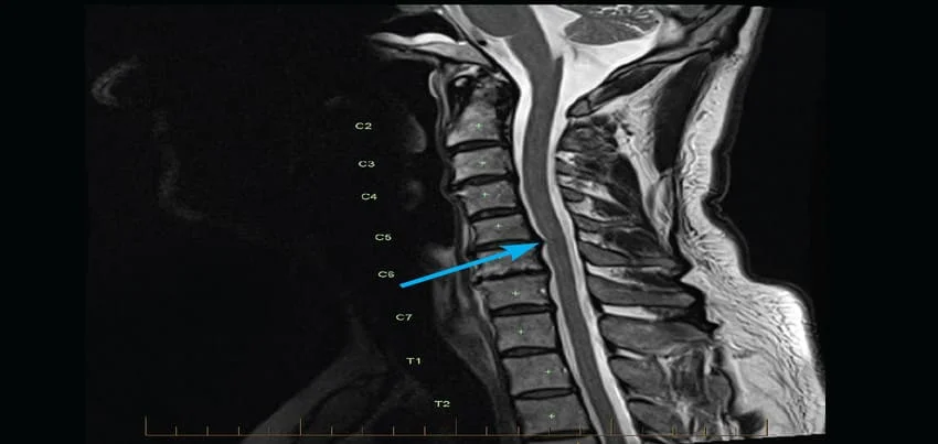
MRI Of The Neck Magnetic Resonance Imaging (MRI) of the neck is a diagnostic test that uses a powerful magnetic field, radio waves, and a computer to produce detailed images of the neck and surrounding structures. The test is conducted to evaluate a range of conditions affecting the neck, including injuries, tumors, infections, and other abnormalities. Test Conducted:During an MRI of the neck, the patient lies on a table that slides into a cylindrical machine that houses the MRI scanner. The scanner uses a magnetic field and radio waves to create images of the neck and surrounding structures. The test typically takes between 30 and 60 minutes to complete, depending on the complexity of the exam and the patient’s ability to remain still during the procedure. Test Preparation:For an MRI of the neck, patients should wear comfortable, loose-fitting clothing without metal zippers or buttons. The patient will be asked to remove any metal objects, such as jewelry, watches, or belts. In some cases, the patient may need to fast for a few hours before the exam, depending on the reason for the test. Common Symptoms for Ordering the Test:MRI of the neck may be ordered by a physician if a patient has symptoms such as neck pain, stiffness, or limited range of motion, difficulty swallowing, hoarseness or other voice changes, or unexplained lumps or bumps in the neck. The test can help diagnose a range of conditions affecting the neck and surrounding structures, including injuries, tumors, infections, and other abnormalities. Time Taken for the Test and Interpretation of Results:An MRI of the neck typically takes between 30 and 60 minutes to complete, and patients can return to their normal activities immediately after the test. The images produced by the MRI are examined by a radiologist, who will interpret the results and provide a report to the ordering physician. The physician will then review the results with the patient and determine the appropriate course of treatment based on the findings. MRI of the neck is an important tool for diagnosing and treating a range of conditions affecting the neck and surrounding structures, and can be used in regular health checkups and corporate wellness programs. Early detection of neck problems is critical to maintaining good health and wellness. In addition, some corporate health programs offer wellness screenings that include neck exams, and an MRI of the neck may be ordered as part of a comprehensive wellness evaluation. In conclusion, MRI of the neck is a valuable diagnostic tool that can help detect a range of neck and surrounding structure conditions that can affect overall health and wellness. The test is non-invasive and typically takes between 30 and 60 minutes to complete, with some preparation required such as removing metal objects and fasting for a few hours before the exam. The results are interpreted by a radiologist and reviewed by the ordering physician, who will determine the appropriate course of treatment based on the findings. Regular health checkups and corporate wellness programs can also incorporate neck exams, including MRI of the neck, to help detect and manage neck problems.

Read More

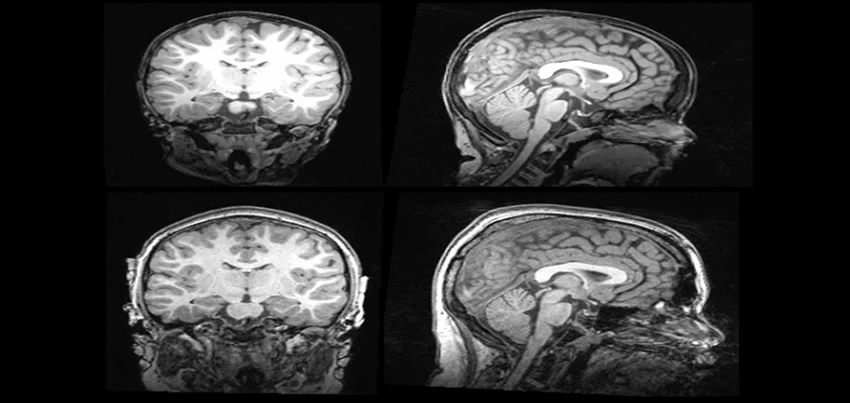
MRI Of The Brain Magnetic Resonance Imaging (MRI) of the brain is a medical imaging test that uses a powerful magnetic field, radio waves, and a computer to create detailed images of the brain and surrounding structures. It is a non-invasive and painless diagnostic test that provides valuable information about the brain’s structure and function. The Test Procedure:During an MRI Brain scan, the patient lies on a table that slides into a tunnel-shaped machine. The patient’s head is secured with straps, and they are provided with earplugs to protect against the loud banging and knocking noises the machine makes during the procedure. The test typically takes between 30 to 60 minutes to complete, depending on the type of MRI and the complexity of the images required. To create clear and detailed images, the patient must remain still during the test. The technician will provide the patient with instructions and may use a communication system to keep in touch with the patient during the procedure. Some types of MRI scans may require a contrast agent injection to enhance the imaging, which is administered through an IV line in the patient’s arm. Test Preparation:Preparation for an MRI Brain scan may vary depending on the type of scan ordered by the physician. Generally, the patient is instructed to avoid wearing any metallic objects, such as jewelry, watches, or clothing with metal zippers, during the procedure. Patients should also inform the technician if they have any implanted medical devices, such as pacemakers, as these may interfere with the MRI machine. Patients may be asked to avoid eating or drinking for several hours before the test, especially if they are undergoing a contrast-enhanced scan. The technician will provide specific instructions to the patient before the test. Common Symptoms for Ordering the Test: An MRI Brain scan may be ordered by a physician for a variety of reasons, including the following symptoms:HeadachesSeizuresDizzinessNumbness or tinglingMemory loss or confusionSuspected brain tumor or other abnormal growthsStroke or other vascular disordersTraumatic brain injuryMultiple sclerosis or other neurological disordersMonitoring of brain function during surgery or treatment Health Checkup, Wellness, and Corporate Health:An MRI Brain scan is often included as part of a comprehensive health checkup or wellness program, especially for individuals at higher risk of neurological disorders. Corporate health programs may also offer MRI Brain scans as a screening tool for employees, especially those who work in high-stress or high-risk environments. The results of an MRI Brain scan may help physicians identify potential health risks and develop personalized treatment plans. In some cases, early detection of neurological disorders can improve the effectiveness of treatment and increase the chances of successful recovery. Interpretation of Results:Interpreting the results of an MRI Brain scan requires specialized training and expertise. The images produced by the MRI machine are highly detailed and may reveal subtle changes in the brain’s structure or function. A radiologist or neurologist typically reviews the images and provides a report to the referring physician. The results of an MRI Brain scan may indicate the presence of abnormal growths, such as tumors or cysts, or changes in brain tissue consistent with neurodegenerative diseases. The images may also reveal vascular abnormalities, such as aneurysms or arteriovenous malformations (AVMs), or evidence of stroke or other cerebrovascular disorders. In some cases, the results of an MRI Brain scan may be inconclusive or require further testing or evaluation. The physician will discuss the results with the patient and develop a treatment plan based on the findings. In conclusion, an MRI Brain scan is a valuable diagnostic tool that provides detailed information about the structure and function of the brain.

Read More

MRI Of The Pelvis Magnetic Resonance Imaging (MRI) of the pelvis is a non-invasive diagnostic test that uses a powerful magnet, radio waves, and a computer to produce detailed images of the organs and tissues in the pelvis. The test is conducted to evaluate the pelvis for various conditions that can cause pain, abnormal bleeding, or other symptoms. Test Conducted:During the MRI of the pelvis, the patient lies on a table that slides into a cylindrical machine that houses the MRI scanner. The scanner uses a magnetic field and radio waves to create images of the pelvis. The test takes between 30 and 60 minutes to complete, depending on the complexity of the exam and the patient’s ability to remain still during the procedure. Test Preparation:For an MRI of the pelvis, patients should wear comfortable, loose-fitting clothing without metal zippers or buttons. The patient will be asked to remove any metal objects, such as jewelry, watches, or belts. The patient may also be asked to fast for a few hours before the exam, depending on the reason for the test. Common Symptoms for Ordering the Test:MRI of the pelvis may be ordered by a physician if a patient has symptoms such as pelvic pain, abnormal bleeding, difficulty urinating or having a bowel movement, or signs of a tumor or other abnormality in the pelvis. The test can help diagnose a range of conditions affecting the pelvis, including ovarian cysts, fibroids, endometriosis, pelvic inflammatory disease, and other abnormalities. Time Taken for the Test and Interpretation of Results:An MRI of the pelvis typically takes between 30 and 60 minutes to complete, and patients can return to their normal activities immediately after the test. The images produced by the MRI are examined by a radiologist, who will interpret the results and provide a report to the ordering physician. The physician will then review the results with the patient and determine the appropriate course of treatment based on the findings. MRI of the pelvis is an important tool for diagnosing and treating a range of conditions that can affect overall health and wellness. Regular health checkups can help detect and manage pelvic problems before they become more serious. Additionally, many corporate health programs offer wellness screenings that include pelvic exams, and an MRI of the pelvis may be ordered as part of a comprehensive wellness evaluation. In conclusion, MRI of the pelvis is a valuable diagnostic tool that can help detect a range of pelvic conditions that can affect overall health and wellness. The test is non-invasive and typically takes between 30 and 60 minutes to complete, with some preparation required such as removing metal objects and fasting for a few hours before the exam. The results are interpreted by a radiologist and reviewed by the ordering physician, who will determine the appropriate course of treatment based on the findings. Regular health checkups and corporate wellness programs can also incorporate pelvic exams, including MRI of the pelvis, to help detect and manage pelvic problems.

Read More

Alcohol Abuse & Alcoholism Alcohol abuse and alcoholism are serious medical conditions that can have a detrimental effect on a person’s physical and mental health, as well as their relationships and overall quality of life. Symptoms of alcohol abuse include drinking more than intended, difficulty controlling drinking habits, problems at work or school, legal issues, and continued drinking despite negative consequences. Alcoholism, also known as alcohol addiction or alcohol dependence, is characterised by a strong craving for alcohol, difficulty controlling drinking, and withdrawal symptoms when not drinking. Diagnosis of alcohol abuse or alcoholism typically involves a physical examination, blood tests, and a psychological evaluation. The most commonly used tool for diagnosing alcohol use disorder is the Alcohol Use Disorders Identification Test (AUDIT). Treatment for alcohol abuse and alcoholism typically involves a combination of therapy and medication. Behavioural therapies, such as cognitive-behavioural therapy and motivational interviewing, can help people learn new coping mechanisms for dealing with stress and triggers for drinking. Medications, such as disulfiram, naltrexone, and acamprosate, can help reduce cravings and prevent relapse. Prevention of alcohol abuse and alcoholism is crucial in reducing the number of individuals who develop the condition. One of the best ways to prevent alcohol abuse is to educate people about the risks associated with heavy drinking. Interventions such as school-based programs, workplace initiatives, and community-wide campaigns can help raise awareness about the dangers of excessive drinking. Diet and exercise are also important in preventing alcohol abuse and alcoholism. Eating a healthy, balanced diet can help reduce cravings for alcohol and promote overall health. Regular exercise can also help reduce stress and improve mood, which can make it easier to resist the temptation to drink. To prevent alcohol abuse and alcoholism, it is important to be mindful of your drinking habits, educate yourself about the risks associated with heavy drinking, and seek help if you think you may have a problem. Additionally, practicing a healthy lifestyle including a balanced diet, regular exercise, and stress-reducing activities can help prevent the onset of alcohol abuse and alcoholism.

Read More

Anxiety Anxiety is a mental health disorder characterized by feelings of worry, nervousness, and fear. These feelings can be overwhelming and interfere with daily activities. Anxiety can manifest in different ways, such as generalized anxiety disorder, panic disorder, social anxiety disorder, and specific phobias. Symptoms of anxiety can include excessive worry and fear, difficulty concentrating, sleep disturbances, irritability, and physical symptoms such as muscle tension, headaches, and stomach upset. To diagnose anxiety, a healthcare provider will typically perform a physical examination and take a medical history. They may also use diagnostic tools such as the Diagnostic and Statistical Manual of Mental Disorders (DSM-5) or the Hamilton Anxiety Rating Scale (HARS) to assess the severity of symptoms and make a diagnosis. Common treatment options for anxiety include therapy, such as cognitive-behavioral therapy (CBT) and talk therapy, and medication, such as antidepressants and anti-anxiety medications. Health check up is a good way to identify the symptoms of anxiety and start the diagnosis process. It is important to schedule regular check-ups to monitor symptoms and progress. To prevent anxiety, it is important to maintain a healthy lifestyle. This includes eating a balanced diet that is rich in fruits, vegetables, and whole grains, and avoiding processed foods and sugary drinks. It’s also important to get enough sleep, exercise regularly, and manage stress through relaxation techniques such as deep breathing, yoga, and meditation. Exercise is a great way to prevent and manage anxiety. Regular exercise can help to reduce stress and tension, improve mood, and promote better sleep. It is recommended to aim for at least 30 minutes of moderate-intensity physical activity on most days of the week. It’s also important to avoid or limit the use of alcohol and caffeine, which can trigger or worsen anxiety symptoms. Building a support system with friends, family, and mental health professionals can also be helpful in managing and preventing anxiety. In conclusion, anxiety is a mental health disorder characterized by excessive worry and fear. It is important to schedule regular health check ups to identify and diagnose anxiety, and to work closely with a healthcare provider to develop an effective treatment plan. A healthy diet, regular exercise, good sleep, and stress management techniques can also help to prevent and manage anxiety.

Read More

Schizophrenia Schizophrenia is a serious mental disorder that affects how a person thinks, feels, and behaves. It is characterized by a wide range of symptoms, including hallucinations, delusions, disordered thinking, and abnormal movements. Symptoms of schizophrenia typically develop in the late teens or early adulthood and can vary greatly from person to person. Common symptoms include hearing voices, seeing things that are not there, having false beliefs, and disorganized thinking. Some people with schizophrenia may also experience difficulty with social interactions, lack of motivation, and apathy. Diagnosis of schizophrenia typically involves a combination of clinical evaluation, psychological tests, and imaging studies. A clinical evaluation typically includes a thorough physical and mental health examination, as well as a review of the patient’s medical and psychiatric history. Psychological tests, such as the Positive and Negative Syndrome Scale (PANSS), can also be used to assess the severity of symptoms. Imaging studies, such as magnetic resonance imaging (MRI) or computed tomography (CT) scans, can be used to rule out any underlying physical conditions that may be contributing to the symptoms. Treatment for schizophrenia typically involves a combination of medication and psychotherapy. Antipsychotic medications, such as haloperidol and risperidone, are commonly used to reduce symptoms such as hallucinations and delusions. These medications can be effective in reducing symptoms, but they can also have serious side effects, such as weight gain and diabetes. Psychotherapy, such as cognitive-behavioral therapy (CBT), can also be helpful in addressing the emotional and social difficulties that often accompany schizophrenia. Preventing the onset of schizophrenia is difficult, as the exact cause of the disorder is not yet fully understood. However, some researchers believe that a combination of genetic and environmental factors may play a role. Studies have shown that certain environmental factors, such as prenatal exposure to viruses or toxins, can increase the risk of developing schizophrenia. Annual health check-ups and corporate health and wellness programs can play a role in the prevention of schizophrenia by identifying and addressing risk factors early on. These check-ups can include assessments of mental health as well as physical health and can help detect any early warning signs of schizophrenia. Corporate health and wellness programs can provide education and resources on mental health and wellness, and can also create a supportive work environment for those who may be at risk for developing schizophrenia. Diet and exercise can also play a role in preventing the onset of schizophrenia. Eating a healthy and balanced diet, rich in fruits, vegetables, and lean proteins, can help to reduce the risk of developing the disorder. Regular physical activity, such as walking, running, cycling, or swimming, can also help to reduce the risk of developing schizophrenia. In summary, Schizophrenia is a serious mental disorder that affects how a person thinks, feels and behaves, characterized by symptoms such as hallucinations, delusions and disordered thinking. Its diagnosis involves a combination of clinical evaluation, psychological tests, and imaging studies. Its treatment typically involves a combination of medication and psychotherapy. Preventing its onset is difficult, but studies have shown that some environmental factors and genetic may play a role. Annual health check-ups, corporate health and wellness programs and a healthy diet and regular exercise can play a role in preventing the onset of schizophrenia.

Read More

MRI Brain Screening Magnetic Resonance Imaging (MRI) is a non-invasive diagnostic tool used to create detailed images of internal organs, tissues, and structures of the body. MRI Brain Screening is a specialised imaging procedure used to evaluate the structures and tissues of the brain. The procedure is conducted by a trained radiologic technologist, who will help you lie down on a flat table that slides into the MRI machine. The machine uses a strong magnetic field and radio waves to create high-resolution images of the internal structures of the brain. During the procedure, you will need to remain still, and may be asked to hold your breath for short periods of time. Before the test, you will need to remove any metal objects, such as jewellery or watches, as these can interfere with the magnetic field. In some cases, you may be given a contrast agent, such as gadolinium, through an intravenous (IV) injection to enhance the visibility of certain structures. There are various symptoms and conditions that may warrant an MRI Brain Screening, including headaches, seizures, dizziness, changes in vision or hearing, and suspected brain tumors or other abnormalities. Additionally, an MRI Brain Screening may be ordered as part of a routine health checkup or wellness program to screen for any underlying conditions that may not be detected through other diagnostic tests. The length of time for an MRI Brain Screening can vary, depending on the complexity of the images required, but typically takes around 30-60 minutes. After the test, the images will be analyzed by a radiologist, who will interpret the results and provide a report to your healthcare provider. The results of an MRI Brain Screening can provide detailed information about the structures and tissues of the brain, including any abnormalities or injuries. The images can be used to diagnose a range of conditions, such as brain tumors, aneurysms, or stroke, and can help guide treatment decisions. In addition to its diagnostic uses, an MRI Brain Screening can also be used in corporate health and wellness programs to screen for underlying conditions that may affect an employee’s health and wellbeing. By identifying potential health issues early, employers can help their employees get the care they need and improve overall productivity and job satisfaction. To prepare for an MRI Brain Screening, you should inform your healthcare provider if you have any metal implants or devices in your body, as these may be affected by the magnetic field. You may also be asked to fast for a period of time before the test, and should avoid wearing any metal objects, such as jewelry or watches. In summary, MRI Brain Screening is a non-invasive diagnostic tool that uses magnetic fields and radio waves to create high-resolution images of the internal structures of the brain. It is a safe and effective way to diagnose a range of conditions, including brain tumors, aneurysms, or stroke. The test preparation typically involves removing metal objects and, in some cases, receiving a contrast agent through an IV injection. The procedure can take 30-60 minutes, and the results are interpreted by a radiologist who provides a report to your healthcare provider. MRI Brain Screening can be used in corporate health and wellness programs to screen for underlying conditions that may affect an employee’s health and wellbeing, and help identify potential health issues early. MRI: Brain Screening Magnetic Resonance Imaging (MRI) is a powerful diagnostic tool used for detailed imaging of the brain and other soft tissues. It is non-invasive, uses no ionizing radiation, and provides high-resolution images that are crucial for diagnosing and monitoring various neurological conditions. What is a Brain MRI? A Brain MRI uses strong magnetic fields and radio waves to produce detailed images of the brain and its structures. It can be used to screen for a wide range of conditions, including structural abnormalities, tumors, inflammation, vascular issues, and neurodegenerative diseases. Why is a Brain MRI Done? A Brain MRI is performed for several reasons, including: 1. Diagnostic Purposes:o Headaches: To identify underlying causes like tumors or vascular abnormalities.o Seizures: To detect structural abnormalities or lesions.o Stroke: To visualize blood flow issues and brain tissue damage.o Trauma: To assess injuries and bleeding.o Dementia: To evaluate changes associated with neurodegenerative diseases like Alzheimer’s.o Infections: To identify brain infections or abscesses.o Multiple Sclerosis (MS): To detect and monitor demyelination.2. Pre-Surgical Planning:o Detailed imaging helps surgeons plan procedures involving the brain, minimizing risks and improving outcomes.3. Follow-Up and Monitoring:o For ongoing conditions or post-treatment evaluation to check for recurrence or progression.4. Screening for Asymptomatic Conditions:o In individuals with high risk factors or family history of neurological diseases. Types of Brain MRI Sequences MRI uses various sequences to highlight different tissues and abnormalities in the brain: 1. T1-Weighted Imaging:o Good for visualizing normal brain anatomy.o CSF (Cerebrospinal Fluid) appears dark, and fat (in myelin) appears bright.2. T2-Weighted Imaging:o Excellent for detecting lesions and abnormalities.o CSF appears bright, making it useful for identifying fluid-related issues like edema.3. FLAIR (Fluid-Attenuated Inversion Recovery):o Similar to T2 but with suppressed fluid signals.o Ideal for detecting lesions near the ventricles or in the cortical and subcortical areas.4. DWI (Diffusion-Weighted Imaging):o Detects changes in the movement of water molecules in tissue.o Crucial for early detection of stroke and acute ischemia.5. SWI (Susceptibility-Weighted Imaging):o Sensitive to blood products, calcium, and iron.o Useful for detecting microbleeds and vascular malformations.6. Contrast-Enhanced MRI:o Uses gadolinium-based contrast agents to highlight blood vessels and areas of disruption in the blood-brain barrier.o Enhances the visibility of tumors, inflammation, and infections.7. Functional MRI (fMRI):o Measures brain activity by detecting changes associated with blood flow.o Useful for pre-surgical planning and studying brain function. Preparation for a Brain MRI 1. Safety Screening:o Ensure no contraindications for MRI, such as implanted metallic devices or foreign bodies, which can interact with the magnetic field.o Patients with certain types of pacemakers, aneurysm clips, or cochlear implants may not be eligible for MRI.2. Fasting:o Usually not required unless specified for specific procedures or contrast studies.3. Clothing

Read More
1 2 3 38

Get A Quote


    Request Callback From Health Advisor

    4.7

    Based on 423

    google

    Reviews

    Why Choose PrognoHealth..??

    • Best health services from different health service providers offered under one umbrella.

    • Big savings on your healthcare cost

    • Choice & convenience to your employees to avail health services at any of our network centers

    • Customized Health Packages

      Request Callback From Health Advisor

      4.7

      Based on 423

      google

      Reviews

      Why Choose PrognoHealth..??

      • Best health services from different health service providers offered under one umbrella.

      • Big savings on your healthcare cost

      • Choice & convenience to your employees to avail health services at any of our network centers

      • Customized Health Packages